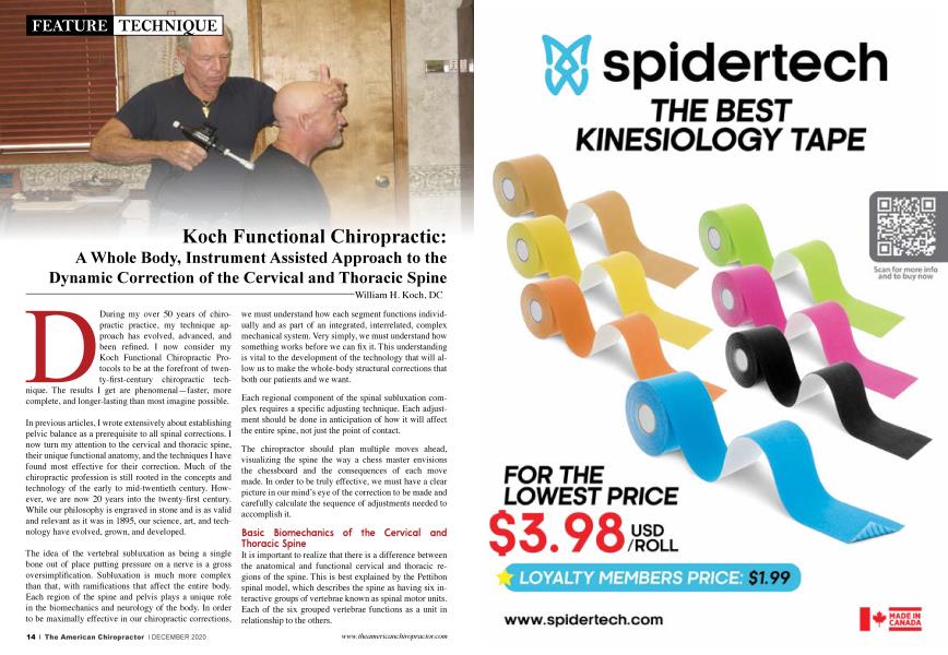
During my over 50 years of chiropractic practice, my technique approach has evolved, advanced, and been refined. I now consider my Koch Functional Chiropractic Protocols to be at the forefront of twenty-first-century chiropractic techniques. The results I get are phenomenal—faster, more complete, and longer-lasting than most imagine possible.
In previous articles, I wrote extensively about establishing pelvic balance as a prerequisite to all spinal corrections. I now turn my attention to the cervical and thoracic spine, their unique functional anatomy, and the techniques I have found most effective for their correction. Much of the chiropractic profession is still rooted in the concepts and technology of the early to mid-twentieth century. However, we are now 20 years into the twenty-first century. While our philosophy is engraved in stone and is as valid and relevant as it was in 1895, our science, art, and technology have evolved, grown, and developed.
The idea of the vertebral subluxation as being a single bone out of place putting pressure on a nerve is a gross oversimplification. Subluxation is much more complex than that, with ramifications that affect the entire body. Each region of the spine and pelvis plays a unique role in the biomechanics and neurology of the body. In order to be maximally effective in our chiropractic corrections, we must understand how each segment functions individually and as part of an integrated, interrelated, complex mechanical system. Very simply, we must understand how something works before we can fix it. This understanding is vital to the development of the technology that will allow us to make the whole-body structural corrections that both our patients and we want.
Each regional component of the spinal subluxation complex requires a specific adjusting technique. Each adjustment should be done in anticipation of how it will affect the entire spine, not just the point of contact.
The chiropractor should plan multiple moves ahead, visualizing the spine the way a chess master envisions the chessboard and the consequences of each move made. In order to be truly effective, we must have a clear picture in our mind's eye of the correction to be made and carefully calculate the sequence of adjustments needed to accomplish it.
Basic Biomechanics of the Cervical and Thoracic Spine
It is important to realize that there is a difference between the anatomical and functional cervical and thoracic regions of the spine. This is best explained by the Pettibon spinal model, which describes the spine as having six interactive groups of vertebrae known as spinal motor units. Each of the six grouped vertebrae functions as a unit in relationship to the others.
The six spinal motor units are:
The cranial-cervical complex is at the top of the neurological and biomechanical hierarchy. While the anatomical cervical spine consists of C1-C7 vertebrae, the functional cervical spine consists of spinal units one and two, which are the skull and atlas, and C2-C5, with the fifth cervical being at the apex of the normal cervical lordotic curve.
The functional thoracic spine begins at the sixth cervical vertebrae and extends down to the second lumbar vertebrae, the apex of the normal lumbar lordotic curve.
The apices of the cervical and lumbar lordosis are pivot points or fulcrums that are critical to spinal motion and shock absorption, providing a mechanical advantage for the musculature to work with maximum efficiency and minimal energy expenditure.
Understanding the functional relationship between the cervical and thoracic spine is the key to effective correction of the cervical component of the subluxation complex.
The thoracic region includes the T spine, the attached rib cage, and the organs within (heart, lungs, and liver) and makes up approximately 60% of the mass of the body. Its sheer size and weight make the thoracic region the functional base against which the cranial-cervical and lumbar-pelvic motor units work. This was discovered in the early days of the NASA space program when the astronauts were observed moving about in a weightless environment.
NASA scientists observed this thoracic region as the stable base connecting the balancing and counterbalancing mechanisms of the cranial-cervical and lumbar-pelvic opposing gimbals. Under the influence of Earth’s gravity, these complex coupled joints are critical to the neurological self-righting reflex that allows us to walk upright with balance, strength, and agility. This same system of gimbals allows us to compensate for spinal imbalances caused by subluxation and decompensate when subluxations are corrected by good chiropractic adjustments
The engineering of the thoracic region of the spine is unique because it is designed to deflect direct force away from the core of the body in order to protect the vital organs from injury. Because of this design characteristic, the thoracic spine repels and dissipates the posterior-to-anterior (P to A) adjustments commonly used by many chiropractors. Although well intended, P to A adjustments in the thoracic spine not only are ineffective and biomechanically incorrect but they also can and often do radiate forces into the lower cervical and upper lumbar pivotal vertebrae, adversely affecting their alignment by a whiplash-like action. This kind of unintended consequence only serves to complicate treatment.
The adjustment of the thoracic spine is an important component of a complete spinal correction, but it must be done in a way that the body can accept rather than reject. Successful biomechanically correct adjustments of the T spine are done A to P with the patient supine or standing. The force is directed up the plane of the ribs and toward a fulcrum provided by the doctor’s hand placed at the point of the spine where a sheer force or movement is desired. This thrust is applied at the bottom of the patient's expiration for the best result. However, another technique approach is easier on the doctor and the patient. I will discuss that alternative that utilizes the handheld VibraCussor and ArthroStim instruments later in this article.
The thoracic spine is the least mobile region of the spine due to the attachment of the twelve pairs of ribs to the vertebrae posteriorly and the sternum, via the costal cartilages anteriorly. The greatest freedom of movement is the flexion and extension, which occurs in conjunction with the expansion and contraction of the rib cage during the respiratory cycle of inhalation and expiration. This same cycle of physiological movement is an integral part of the primary sacral respiratory motion, which propels the cerebrospinal fluid up, down, and around the spinal cord and brain. That makes the movement of the thoracic spine doubly critical to life and health.
Fixation is the primary thoracic spinal malfunction. It occurs in response to lumbar misalignment and pelvic imbalance from below and cranial-cervical misalignment from above. Thoracic fixation not only inhibits the fullness of the breathing cycle, but also prevents the normal, smooth, and free interaction between the cranial-cervical and lumbar-pelvic gimbals. The consequence of this is loss of overall spinal motion, resiliency, and shock absorption. Very importantly, it also creates a resistance to the correction of the subluxation components above and below it.
This makes the mobilization to the thoracic spine critical to the corrective process. When we fail to integrate the mobilization of the thoracic spine with our adjustments of the other areas of the spine, those corrections are doomed to fail. Therein lies one of the chief complaints among chiropractic patients —“adjustments do not hold,” necessitating a seemingly endless series of office visits for readjustment. When adjustments do not hold, it frustrates and discourages patients, creating conflict between the doctor, patient, and insurance companies.
The obvious solution is better adjustments that hold. First, it is important to understand the basic physics of the subluxation complex, which is expressed by the following simple equation: The degree of spinal misalignment X the degree of resistance holding it = the force needed to correct it. Therefore, the reduction of the resistance component is the logical first step toward effective, lasting subluxation correction.
Most chiropractors recognize the importance of “loosening up the spine” in preparation for an adjustment by reducing muscle spasms. Typically, they use such things as hot packs, massage, or an electronic modality, all of which are marginally effective, providing only temporary relief at best.
The reduction of subluxation resistance is best accomplished by the restoration of intersegmental spinal motion that had been locked by muscle splinting and guarding directed by the motor cortex. This is a normal defensive sensory-motor reaction caused by the firing of joint and muscle mechanoreceptors when they detect tissue stress, injury, and inflammation.
Much of the chronic pain and restricted spinal and extremity motion we see in our patients is the result of the continued firing of the mechanoreceptors long after the guarding and splinting spasms are needed to protect an injured area.
The exciting news is that advanced adjusting techniques using the ArthroStim and VibraCussor instruments by Impac, Inc. allow us to reset the mechanoreceptors, which send updated status reports to the brain. The brain then revises its map. The ability of the brain to reprogram and revise its mapping of the body is known as neuroplasticity. When the brain map no longer includes a report of an injury, muscle splinting is not needed, spasm is eliminated, and so is resistance to subluxation correction. The body may now freely accept adjustments and allow them to “hold.”
Twenty-first-century chiropractic instrument-adjusting techniques allow us to utilize the somatosensory and motor functions of the body and nervous system to orchestrate structural corrections better, faster, more comfortably, and with longer-lasting results than was possible with conventional manual adjusting techniques.
The most serious and stubborn resistance to cervical correction is in the upper thoracic segments T1-T4. This block of vertebrae is the least movable in the spine, with T1 being the least movable. T1 is the posterior anchor point for the cervical erector muscles and the supraspinous ligament, which is a continuation of the ligamentum nuchae that attaches to the external occipital protuberance (EOP) of the skull and the spinous processes of the cervicals. It continues inferiorly attaching to the spinous process of all thoracic and lumbar vertebrae, ultimately attaching to the posterior sacral tubercles and blending into the neighboring fascia.
The significance of this is that T1 and the spine inferior to it are under constant stress while we engage in any activity when the head is down and the neck is in forward flexion. That means almost everyone spends much of their day with their spine under excess tension. The position called “tech neck” is the best example of the posture assumed while using electronic devices (computers, phones, and tablets). The result is the chronic anterior head translation that so many people now have. It is the reason so many people now suffer from neck, upper back, and shoulder tension and pain, headaches, and cervical/brachial radiculopathies. This chronic anterior translation of the head causes an accelerated development of degenerative joint disease and disc narrowing at C5/C6 as well as the dreaded dowager's hump that so many women and some men develop.
The combination of anterior head translation and thoracic fixation can inhibit breathing mechanisms, causing up to a 30% reduction of vital lung capacity. In my experience with several patients diagnosed with COPD who required oxygen concentrators to maintain blood oxygen levels, they were able to breathe well and maintain blood oxygenation without the concentrator after successful reduction of anterior head translation and thoracic fixation. Several important mechanisms of neurological stress also are caused by subluxation of the skull-atlas and C2-C5 motor units relative to the upper thoracic C6-T7 motor unit. It is important that every DC fully appreciate and understand these nerve-pressure and tension-applying mechanisms and the steps necessary to reduce them.
The benefits of restoring motion to a previously misaligned and fixated spine are tremendous. Among the mechanoreceptors we are resetting with the ArthroStim instrument are the type one, two, and three. Besides providing the sensory cortex with a cascade of new information, causing it to create a revised brain map or picture of the body, the type four nociceptive-pain, and alarm-related mechanoreceptors are inhibited.
Because pain and other nociceptive input to the brain trigger the sympathetic portion of the autonomic nervous system, reduction of the nociceptive input suppresses the sympathetic, allowing the reestablishment of normal parasympathetic dominance. In other words, it takes us out of “fight-or-flight” and back to “rest-and-relax.” Restoration of the normal autonomic nervous system balance is necessary for healing on every level.
Loss of the cervical lordotic curve is an almost universal spinal misalignment. It has the effect of increasing spinal cord tension by as much as 24%. The resulting extrusion of the dura can apply as much as 30 pounds per square inch of pressure to the spinal cord. This excessive cord tension can pull the medulla and brain stem down too tightly against the floor of the cranium, especially in the area of the foramen magnum.
Upper cervical subluxations involving the occiput, atlas, and axis put serious pressure on the brain stem, which extends through the foramen magnum, the ring of the atlas, and down to the inferior aspect of the neural canal of the axis. Most cervical subluxations have a rotational component. Those that involve counter-rotation between atlas and axis add a complicating factor and are usually associated with ligament and other soft tissue injuries, posing an additional challenge to the DC.
Few would argue that conventional chiropractic techniques are done mainly with the patient lying still in the prone position. (I have heard several practice management consultants actually say that the only part of the chiropractor the patient should ever see after the first visit is his shoes, viewed through the facepiece of a hi-lo table.) Let me say here and now that my views about patient care, practice management, and technique are totally contrary to most of the practice management gums. If this is not already obvious, it soon will be.
With conventional chiropractic techniques, the patient lies still on the adjusting table, taking a passive role. With Koch Functional Chiropractic Protocols, the patient plays an active role and is directed by the doctor through various movements of the spine and extremities during the corrections. Active participation creates a dramatically different experience for patients. They feel a greater connection when actively working with their doctor, and, most importantly, patients experience an immediate improvement in body balance, strength, and range of motion. Positive outcomes are then objectively confirmed via post-correction muscle testing.
Rather than static spine-only adjusting of earlier chiropractic techniques, Koch Functional Chiropractic Protocols offer an innovative, dynamic, whole-body approach with an organized system of protocols that represent much of the best of twenty-first-century chiropractic. Now that I have given you a new vision and the rationale behind this departure from traditional bone-moving chiropractic, I will describe the actual adjusting techniques.
A Twenty-First-Century Approach to Thoracic and Cervical Corrections
I begin cervical corrections by mobilizing the thoracic spine to reduce fixations and normalize intersegmental motion. This is accomplished quickly and comfortably by using the VibraCussor and ArthroStim instruments by Impac, Inc.
With the patient seated on the adjusting table, the doctor contacts the sacral base with the soft padded VibraCussor head. The instrument is set on a medium frequency, and the patient is instructed to rotate their head and neck through the complete right-and-left rotatory range of motion. It takes only a few moments before both doctor and patient notice a definite improvement in that range of motion.
Next, while the doctor continues to percuss the sacral base, the patient is instructed to exhale fully as they bend forward at the waist with the head and neck in flexion. The patient is then told to inhale as they move from flexion to full spinal extension. This is repeated two to three times while the doctor continues to percuss the sacral base.
This simple, comfortable, and non-threatening procedure is remarkably effective at releasing fixations throughout the spine and can be used even on frail, timid, or frightened patients.
The procedure works by utilizing the engineering design of the spine in which the 24 vertebrae decrease in size and density from L5 to the atlas. When the wave of percussive energy is introduced to the sacral base, it radiates up the spine, amplifying and accelerating as the vertebrae reduce in size. The combined cervical rotation and spinal flexion/extension motion with respiration in conjunction with the action of the VibraCussor are highly effective in reducing spinal fixation and increasing intersegmental motion.
Then the doctor switches to the ArthroStim instrument, fitted with a bifurcated gliding sleeve. With the instrument set at its maximum of twelve toggle-recoils per second, the doctor glides the ArthroStim instrument from L5 through the upper cervical spine using a gentle level of pressure that is comfortable to the patient. After one or two passes of the instrument, again ask the patient to flex and extend their cervical spine through the complete range of motion while you continue to glide the ArthroStim from the upper thoracic through the top of the cervical spine. Since T1 is strategically important to the corrective process because of the extreme fixation that occurs there, it is beneficial to spend some extra time at that segment.
Also, because there is often the rotation of T1 (as in a Gonstead PLS or PRS listing), the ArthroStim can be used to adjust it accordingly using the appropriate line of drive. To the casual observer, this procedure might appear to be using the ArthroStim to “jackhammer” the bones into place, but nothing could be further from the truth. The objective is not to move vertebrae in the traditional sense. The aberrant afferent input is, instead, a resetting of the joint mechanoreceptors, which sends a cascade of updated, revised information to the sensory cortex, resulting in a revised brain map that reflects a correcting, rather than a subluxated spine. This information is relayed to the motor cortex, which facilitates the correction. This is twenty-first-century chiropractic.
At this point, the doctor should palpate the cervical spine to check for any remaining subluxations. The good news is that if we have established a solid, stable pelvic foundation and adequately mobilized the spine, the powerful self-righting reflex will already have begun to decompensate and disengage, making any cervical subluxations easier to correct. The body will welcome rather than resist the adjustment.
Specific Instrument-Assisted Cervical Corrective Procedure
After T1 correction, what remains will usually be some combination of occipital, atlas, and axis misalignments. In most cases, there will be a left or right rotation of 2C (as in Gonstead PLS or PRS listing). My preferred technique for these cervical corrections (atlas and axis) is using the ArthroStim instrument fitted with a single round (ball tip) or small straight flat-tipped adjusting sleeve.
In the case of a 2C PRS, the procedure would be as follows:
The patient is instructed to first turn their head as far as is comfortable to the left. Next, the patient turns it slowly from left to right against resistance provided by the doctor who applies adjusting thrusts with the instrument in a P to A and right-to-left line of the drive at the right lamina pedicle junction of 2C. This process can be repeated up to three times. Reverse procedure for 2CPLS.
If the doctor decides that the atlas needs to be adjusted, it can be done with the ArthroStim using the small straight sleeve. Position the tip of the instrument on the transverse process of the atlas, direct the line of the drive as appropriate to move the atlas either A to P or P to A (ASRA, ASRP, ASLA, or ASLP). If head tilt or upper cervical fixation persists, a manual occipital adjustment is indicated. I do this as an occipital lift, which incorporates cervical decompression, slight rotation, and a scooping motion. This is a very technique-intensive adjustment that requires training and practice to bring all three force components together at a precise instant. This adjustment has the greatest potential to relieve the most serious brain stem or spinal cord pressure. Learning and perfecting this adjustment is worth any effort it takes.
Chiropractic is a thinking person’s profession. The master chiropractor learns to think three-dimensionally and strategically, like the aforementioned chess master. Always consider the patient holistically and functionally, evaluating, balancing, and tuning up the entire body.
Have a plan and take a systematic approach. Think ahead. Visualize what you want to achieve and how you will do it. Think three-dimensionally. Observe your objective indicators before and after each adjustment because they will help you and the patient know when you have achieved a desired result. Always record what you do in detail because not only will it put you in a very strong medical-legal position to justify your treatment protocols, but it also assists you greatly as you review pre and post-treatment findings before your next session.
This methodical and interactive approach to patient care and record-keeping will pay off in ways you might not expect. It allows you to consistently provide the highest quality of individualized care for your patients and gives the patients a very high level of confidence in you, their doctor. As many of my patients have said, when they feel “this is the kind of care I have been looking for from a doctor,” they eagerly refer their friends and family members. Providing this kind of personalized, exceptional care is the best practice-building tool for which you could hope.
Dr. William H. Koch is a 1967 Cum Laude graduate of Palmer College of Chiropractic in Davenport, Iowa. He practiced in the Hamptons of Eastern Long Island, New York for 30 years and in the Bahamas for 15 years aboard his motor yacht, The Coastal Chiropractor. He is licensed to practice in New York, Florida, and The Bahamas and currently splits his time between Abaco in The Bahamas and his newest practice in Mount Dora, Florida. Now, wanting to give back to the profession he loves, he offers courses on "The Koch Protocols for Integrated, Advanced, Chiropractic Techniques." Simple, Effective, No-Nonsense, and Hands-On. He may be reached by email at [email protected] or DrWilliamHKoch.com.