Arches and Orthotic Support
ORTHOTICS
Mark Charrette
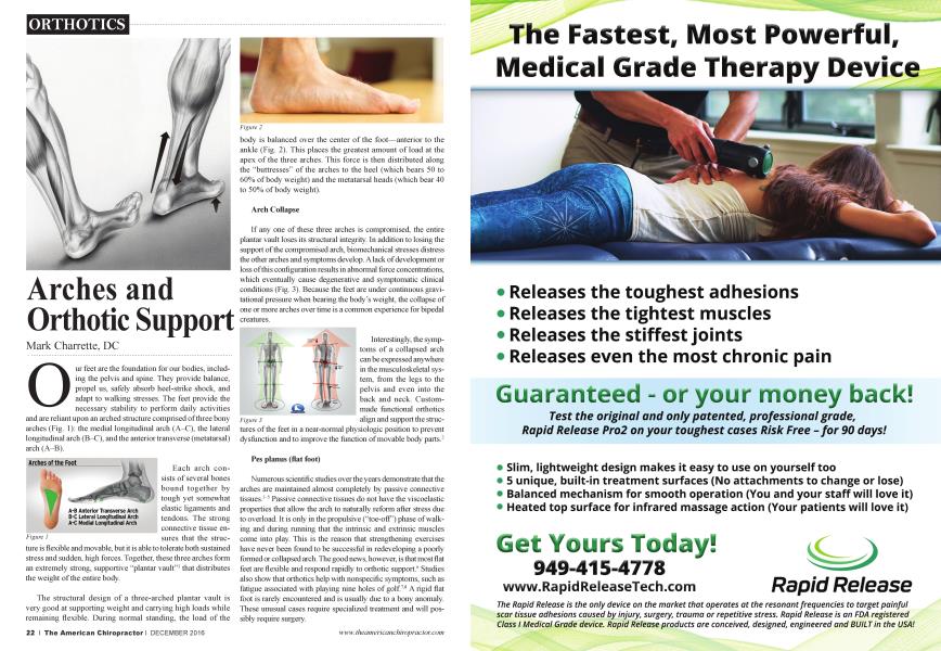
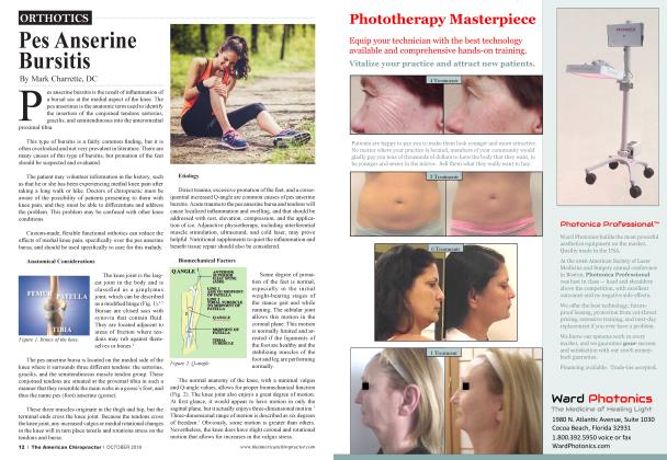
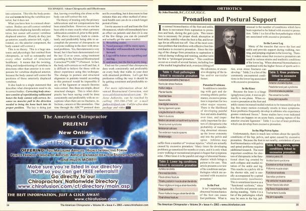
Our feet are the foundation for our bodies, including the pelvis and spine. They provide balance, propel us, safely absorb heel-strike shock, and adapt to walking stresses. The feet provide the necessary stability to perform daily activities and are reliant upon an arched structure comprised of three bony arches (Fig. 1): the medial longitudinal arch (A—C), the lateral longitudinal arch (B—C), and the anterior transverse (metatarsal) arch (A—B).
Each arch consists of several bones bound together by tough yet somewhat elastic ligaments and tendons. The strong connective tissue ensures that the struc-
ture is flexible and movable, but it is able to tolerate both sustained stress and sudden, high forces. Together, these three arches form an extremely strong, supportive “plantar vault”1 that distributes the weight of the entire body.
The structural design of a three-arched plantar vault is very good at supporting weight and carrying high loads while remaining flexible. During normal standing, the load of the
body is balanced over the center of the foot—anterior to the ankle (Fig. 2). This places the greatest amount of load at the apex of the three arches. This force is then distributed along the “buttresses” of the arches to the heel (which bears 50 to 60% of body weight) and the metatarsal heads (which bear 40 to 50% of body weight).
Arch Collapse
If any one of these three arches is compromised, the entir e plantai' vault loses its structural integrity. In addition to losing the support of tiie compromised arch, biomechanical stresses distress tiie other arches and symptoms develop. A lack of development or loss of this configuration results in abnormal force concentrations, which eventually cause degenerative and symptomatic clinical conditions (Fig. 3). Because tiie feet aie under continuous gravitational pressure when bearing the body’s weight, tiie collapse of one or more arches over time is a common experience for bipedal creatures.
Interestingly, the symptoms of a collapsed arch can be expressed anywhere in the musculoskeletal system, from the legs to the pelvis and even into the back and neck. Custommade functional orthotics align and support the struc-
tures of the feet hi a near-normal physiologic position to prevent dysfunction and to improve the function of movable body parts.2
Pes planus (flat foot)
Numerous scientific studies over the years demonstrate that the arches aie maintained ahnost completely by passive connective tissues.3 5 Passive connective tissues do not have the viscoelastic properties that allow tiie arch to naturally reform after stress due to overload. It is only in the propulsive (“toe-off’) phase of walking and during running that the intrinsic and extrinsic muscles come into play. This is the reason that strengthening exercises have never been found to be successful in redeveloping a poorly formed or collapsed arch. The good news, however, is that most flat feet aie flexible and respond rapidly to orthotic support.6 Studies also show that orthotics help with nonspecific symptoms, such as fatigue associated with playing nine holes of golf.78 A rigid flat foot is rarely encountered and is usually due to a bony anomaly. These unusual cases require specialized treatment and will possibly require surgery.
“if any one of these three arches is compromised, the entire plantar vault loses its structural integrity. In addition to losing the support of the compromised arch, biomechanical stresses distress the other arches and
symptoms develop. 5 5
Excessive pronation
At heel strike and dining the initial part of stance phase, the foot normally pronates. This absorbs some of the shock of heel strike and accommodates uneven terrain.
However, if the foot stays in pronation beyond heel strike, it is hyperpronating or going mto prolonged pronation. This movement occurs primarily at the subtalar and talonavicular joints, with excessive loading affecting all of the arches, but the medial arch most acutely. Excessive pronation causes an obvious flattening of the medial longitudinal arch, with a medial and inferior movement of tlie navicular bone. This arch collapse destroys the structural support of the plantai' vault, making the body at risk for subluxations as the musculoskeletal system attempts to adapt and compensate.
How You Can Measure Arch Collapse
You can perform a quick test that measures the change in position of the navicular prominence to quantify tlie presence of arch collapse duiing weight bearing.9 The navicular drop test is especially useful10 because it shows the change in arch height from non-weight bearing to weight bearing, as well as any asymmetry between left and right arches. The test helps to verity poor spinal support from the arches and demonstrates the need for orthotics to the patient.
What Can Orthotics Do?
Static support
During standing posture, the aligmnent of tlie arches in each foot has a significant impact on tlie position of tlie legs and pelvis. When the arches aie low and/or pronating excessively, the lower extremities tend to rotate medially.
Dynamic support
Duiing gait, the foot undergoes substantial changes. The arches and connective tissues must sustain the stress of heel strike, then adapt to the ground during stance phase, and finally become a rigid lever to provide an efficient push-off. This must occur in a coordinated manner, with no glitches or hangups. The foot must permit a smooth transfer of tlie body’s center of mass over the leg to conserve energy and keep the work expenditure at a minimum.11 The heavier a patient is, the greater the stresses on the feet and ankles. This requires an orthotic to be flexible yet supportive. Orthotic designs consider weight and intensity of forces, in addition to encouraging proper movement and function of the foot, while supporting all three arches.
Postural benefits
Because the entire body structure is balanced on one foot at a time when walking and running, improving foot aligmnent can
improve knee, hip, pelvis, and even spinal postural aligmnent. A low femur head seen on properly positioned postural films indicates a difference in leg length. While there are several causes (from injury to growth asymmetry to arch collapse), most patients will benefit from the additional support provided by a pair of orthotics.12 An added heel lift may also be necessary in some cases. Joint degeneration (of the hip, knee, or spinal joints) with wealing of the cartilage requires the additional support and shock absorption provided by orthotics. A pelvic or spinal tilt or recurrent subluxations will often respond rapidly to orthotic support of the arches.13
All It Takes Is a Quick Glance at the Feet
It’s not unusual to have musculoskeletal complaints in the legs, hips, and spine from malfunctioning arches. A brief screening exam can help identify the commonly seen clues. Look for lowered arches, heel eversion, uneven shoe wear, Achilles tendon bowing, and leg length differences. Foot Levelers’ custom-made orthotics can provide much of the support that is lacking and improve locomotive efficiency by guiding the calcaneus and arches through the gait cycle.
References
1. Kapcmdji IA. Physiology of the Joints: Lower Limb (2nded.). New York: Churchill Livingstone, 1981:154-182.
2. Lening PC. Weightbearing casting and orthotics. Digest Chiro Econ 1992;34(5):52.
3. Basmajian JV, Bentzen JW. An electromyographic study of certain
muscles of the leg and foot in the standing position. Surg Gynec & Obstet 1954;98:662-666.
4. Basmajian JV, Stecko G. The role of muscles in arch support of the foot: an electromyographic study. JBone Joint Surg 1963;45A:1184-1190.
5. Huang C-K et al. Biomechanical evaluation of longitudinal arch stability. Foot & Ankle 1993;14:353-357.
6. Kuhn DR, Shibley NJ, Austin WM, Yochum TR. Radiographic evaluation of weight-bearing orthotics and their effect on flexible pes planus. J Manip Physiol Ther 1999;22(4):221-226.
7. Stude DE, Gullickson J. Effects of orthotic intervention and nine holes of simulated golf on dub-head velocity in experienced golfers. J Manip Physiol Ther 2000;23(3):168-174.
8. Stude DE, Gullickson J. Effects of orthotic intervention and nine holes of simulated golf on gait in experienced golfers. J Manip Physiol Ther 2001;24(4):279-287.
9. Gould N. Evaluation of hyperpronation and pes planus in adults. Clin Orthop 1983;181:37-45.
10. Brody D. Techniques in the evaluation and treatment of the ired runner. Orthop Clin North Am 1982;13:541-558.
11. Kirby KA, Biomechanics of the normal and abnormal foot. J Am Podiatr Med Assoc 2000;90:30-34.
12. Baylis WJ, Rzonca EC. Functional and structural limb length discrepancies: evaluation and treatment. Clin Podiatr Med Surg 1988;5:509-520.
13. Rothbart BA, EstabrookL. Excessive pronation: a major biomechanical determinant in the development of chondromalacia and pelvic lists. J Manip Physiol Ther 1988;11:373-379.
Dr Mark Charrette is a 1980 summa cum laude graduate of Palmer College of Chiropractic. He is a frequent guest speaker at Chiropractic colleges worldwide and has taught over fourteen hundred seminars worldwide on |i extremity adjusting, biomechanics, and spinal adjusting * techniques. Contact info: [email protected]