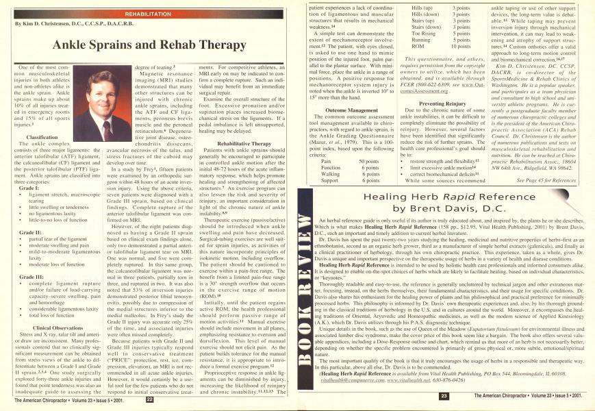
One <>l the mosl common musculoskcletal injuries in both athletes and non-athletes alike is the ankle sprain. Ankle sprains make up about 10% of" all injuries treated in emergency rooms and 15rA of all sports injuries.1 Classification Tlie ankIe comp 1 ex consists of three major ligaments: the anterior talofibular (ATF) ligament, the ealcaneofibular (CF) ligament and the posterior talofibular (PTF) ligament. Ankle sprains are classified into three categories: Grade I: ligament stretch, macroscopic tearing little swelling or tenderness no ligamentous laxity little-to-no loss of function Grade II: partial tear of the ligament moderate swelling and pain mild-to-moderate ligamentous laxity | moderate loss of function ! Grade III: j complete ligament rupture and/or failure of load-carrying capacity-severe swelling, pain and hemorrhage considerable ligamentous laxity total loss of function •j Clinical Observations ; Stress and X-ray, talar lilt and anteri- ! or draw are inconsistent. Many proles- ! sionals contend that no clinically sig- | nificant measurement can be obtained i from stress views of the ankle to dif- { ferentiate between a Grade I and Grade j II sprain.2--*-4 One study surgically I explored forty-three ankle injuries and j found that point tenderness was also an j inadequate guide to assessing the i degree of tearing.5 I Magnetic resonance j imaging (MRI) studies j demonstrated that many j other structures can be ] injured with chronic ankle sprains, including the ATF and CF ligaments, peroneus brevis muscle and the peroneal retinaculum.6 Degenerative joint disease, osteo-chondritis dissecans. avascular necrosis of the talus, and stress fractures of the cuboid may develop overtime. In a study by Frey1, fifteen patients were examined by an orthopedic surgeon within 48 hours of an acute inversion injury. Using the above criteria, seven patients were diagnosed with a Grade III sprain, based on clinical findings. Complete rupture of the anterior talofibular ligament was confirmed on MRI. However, of the eight patients diagnosed as having a Grade II sprain based on clinical exam findings alone, only two demonstrated a partial anterior talofibular ligament tear on MRI. One was normal, and five were completely ruptured. In this same group, the calcaneolibular ligament was normal in three patients, partially torn in three, and ruptured in two. It was also noted that 53'/c of inversion injuries demonstrated posterior tibial tenosyn-ovitis. possibly due to compression of the medial structures inferior to the medial mallcolus. In Frey's study the Grade II injury was accurate only 25% of the lime, and associated injuries were often missed completely. Because patients with Grade II and Grade III injuries typically respond well to conservative treatment ("PRICE": protection, rest, ice. compression, elevation), an MRI is not recommended in all acute ankle injuries. However, it would certainly be a useful tool for the few patients who do not respond to initial conservative treat- ments. For competitive athletes, an MRI early on may be indicated to confirm a complete rupture. Such an individual may benefit from an immediate surgical repair. Examine the overall structure of the foot. Excessive pronation and/or stipulation places increased biome-chanical stress on the ligaments. If a pedal imbalance is left unsupported, healing may be delayed. Rehabilitative Therapy Patients with ankle sprains should generally be encouraged to participate in controlled ankle motion after the initial 4K-72 hours of the acute inflammatory response, which helps promote healing and strengthening of affected structures.7 An exercise program can also lessen the risk and severity of reinjury. an important consideration in light of the chronic nature of ankle instability.s-9 Therapeutic exercise (passive/active) should be introduced when ankle swelling and pain have decreased. Surgical-tubing exercises are well suited for sprain injuries, as activities of this nature incorporate principles of isokinetic motion, including overflow. The patient should be cautioned to exercise within a pain-free range. The benefit from a limited pain-free range is a 30° strength overflow that occurs in the exercise range of motion (ROM).1" Initially, until the patient regains active ROM. the health professional should perform passive range of motion activities.11 Manual exercise should include movement in all planes, emphasizing resistance to eversion and dorsiflexion. This level of manual exercise should not elicit pain. As the patient builds tolerance for the manual resistance, it is appropriate to introduce a formal exercise program.12 Proprioceptive response in ankle ligaments can be diminished by injury, increasing the likelihood of reinjury and chronic instability."-12-1-1 The patient experiences a lack of coordination of ligamentous and muscular structures that results in mechanical weakness.14 A simple test can demonstrate the extent of mechanoreceptor involvement.12 The patient, with eyes closed, is asked to use one hand to mimic position of the injured foot, palm parallel to the plantar surface. With minimal force, place the ankle in a range of positions. A positive response for mechanoreceptor system injury is noted when the ankle is inverted 10° to 15° more than the hand. Outcome Management The common outcome assessment tool management available to chiropractors, with regard to ankle sprain, is the Ankle Grading Questionnaire (Mazur. ct til.. 1979"). This is a 100-point index, based upon the following criteria: Pain 50 points Function 6 points Walking 6 points Support 6 points Hills (up) 3 points Hills (down) 3 points Stairs (up) 3 points Stairs (down) 3 points Toe Rising 5 points Running 5 points ROM 10 points This questionnaire, and others. requires permission from the copyright owners to utilize, which has been obtained, and is available through FCER (800-622-6309. see www.Out-comesAssessment.org. Preventing Reinjury Due to the chronic nature of some ankle instabilities, it can be difficult to completely eliminate the possibility of reinjury. However, several factors have been identified that significantly reduce the risk ol' further sprains. The health care professional's goal should be to: restore strength and flexibility12 limit excessive ankle motion14 correct biomechanical deficits" While some sources recommend ankle taping or use of other support devices, the long-term value is debatable.15 While taping may prevent inversion injury through mechanical intervention, it can may lead to weakening and atrophy of support structures.14 Custom orthotics offer a valid approach to long-term motion control and biomechanical correction.16-17 Kim D. Christensen. DC. CCSP, DACRB. is co-director of the SportsMedicine & Rehab Clinics of Washington. He is a popular speaker, and participates as a team physician and consultant to high school and university athletic programs. He is currently a postgraduate faculty member of numerous chiropractic colleges and is the president of the American Chiropractic Association (ACA) Rehab Council. Dr. Christensen is the author of numerous publications and texts on musculoskeletal rehabilitation and nutrition. He can be reached at Chiropractic Rehabilitation Assoc. 18604 NW 64th Ave.. Ridgefield. WA 98642. See Page 45 for References ...from Page 23 Refer*? nces 1. Frey C. Bell J Teresi L. Kerr R. Fedcr K. A comparison of MRI and clinical examination of acute lateral ankle sprains. Font & Ankle lull 1996: 17:533-537. 2. Bomta PM. Bishop JO. Braly WG. Tullos HS Acute lat- eral ligament injuries: a literature review. Foot & Ankle 1990: 11:107-113. 3. Cox JS. Hewes TF. 'Normal" talar tilt angle. Clin Ortlwp 1979(rclat. res.); 140: 37-41. 4. Frost HM. Hanson CA. Technique for testing the drawer sign in the ankle. Clin Orllmp (rclat. res.I 1977: 123:49-51. 5. Brostrom L. Sprained ankles. Acra Cltir Scand 1965: 130:560-569. 6. Cardone BW. MRI of injury to the lateral collateral liga- mentous complex of the ankle. J Coinput Assist Tmnogr 1993; 17:102-107. 7. Kcrn-Steiner R. Washecheck HS. Kelsey DD. Strategy of exercise prescription using an unloading technique for functional rehabilitation of an athlete with an inversion ankle sprain. J Ortlwp Sports Phys Ther 1999: 29(5):282-287. 8. Roy S. Irvin R. Sports Medicine Prevention. Evaluation. Manusement and Rehabilitation. Englewood Cliffs: Prcnlicc-Hall, I9S3. 9 Holme E. Magnusson SP. Bechcr K. et al. The effect of supervised rehabilitation on strength, postural sway, position sense and re-injury risk after acute ankle ligament sprain. Scaml J Met} Sci Sports 1999; 9(2): 104-109. 10. Davjes GJ. Compendium of lsokinetics in Clinical Usage. UCrosse: S&S Publishers. 1984. 11. Diamond JE. Rehabilitation of ankle sprains. Clinics in SptsMcd 1989:8:877-891. 12 Dcrscheid GL. Broun WC. Rehabilitation of the ankle. Clinics in i;i/.< Med 1985: 4:527-544. 13. Hcncl J. Functional instability following lateral ankle sprain. Sports Med 2(KX): 29(5):361 -371. 14. (Nike Corp.). Lateral ankle sprains. Sport Research Kevin- I989(july/August>. 15. Rclshauge KM. Kilbreath SL. Raymond J. The effect of recurrent ankle inversion sprain and taping on pro-prioception at the ankle. Med Sci Sports Exerc 2000: 32(1):IO-I5. 16. Gross ML. et al. Effccliveness of orthotic shoe inserts in the long-distance runner. Ant J Sprs Med 1991: 19:409-412. 17. Christensen KD. Orthotks: Do They Really Help a Chi- ropritvlH Patient' Roanoke: Foot' Levelers. Inc.. 1990. O