For a clinician, initiating a research study can be highly educational, fun, and validating when you obtain the desired results. It can also be costly, time consuming, and anxiety inducing as you wait for the statisticians to work their dark art. My first foray into research was all of these things, and fortunately, it worked out well at the end of a three-year journey.
In a previous article, we discussed our group’s 2020 study showing that ankle manipulation successfully improved gluteus medius (GMed) activation and force production.1 Our subjects all had a history of ankle sprain and presented with a unilateral hip abductor weakness, suggesting that ankle manipulation can help alleviate GMed arthrogenic muscle inhibition (AMI). AMI is defined as an ongoing reflex inhibition of muscle(s) surrounding a joint following distention or damage [or subluxation] of that joint, resulting in decreased motor neuron pool excitability.2
In addition to being a fairly intuitive concept, alterations of muscle firing secondary to joint dysfunction have been shown repeatedly in literature.3A5-6 I initiated the hip abductor study to help validate its less understood, distal-to-proximal connection with the ankle. (You can find more information on this on my YouTube channel, including a video on diagnosing and treating ankle-induced GMed inhibition.)
Currently, our group is in the data-collection phase of our next study regarding another poorly understood connection: between the thoracic spine and hip adductors. Unfortunately, with the addition of COVID-related delays, I won’t be able to share the results of this study for likely another two years.
In the meantime, we’ll discuss another important but often overlooked manifestation of AMI — inhibition of the psoas muscle. While the role of psoas tightness is well known (i.e., increasing lumbar lordosis and compressing the posterior elements), the effects of psoas weakness are much less commonly discussed, and, despite its importance, there is almost no talk of psoas inhibition.
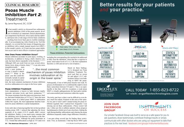
Why does psoas inhibition matter? The answer lies in its three distinct actions. Psoas is best known as a hip flexor,7 but also contributes to lower spine8 91011 and hip joint12 stability. Consequently, any loss of its ability to activate and produce force translates to decreased hip flexion and decreased spine and hip stability. These varied functions are all biomechanically essential. Consequently, psoas inhibition can affect your patients in numerous ways, even when they are doing appropriate core and hip strengthening exercises (as inhibited muscles don’t respond normally to exercise.)
Hip Flexion
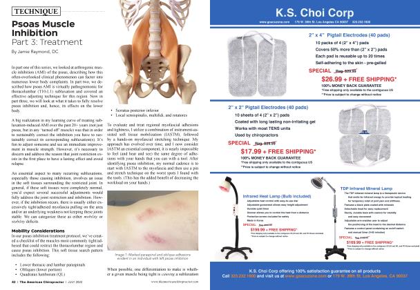
When a muscle is inhibited, neighboring muscles with similar actions frequently become overworked via synergistic dominance. Selective psoas inhibition often characteristically leads to tightness of the other hip flexors, including the iliacus, rectus femoris, sartorius, and TFL. This can then factor into, for example, proximal thigh strains in football players or sprinters, anterior knee pain at the distal rectus femoris in distance runners, or medial knee pain at the distal sartorius in soccer players. This synergist tightness may also limit hip extension, which can then stress the posterior chain.
Spinal Stability
The core region has been described as a compression cylinder, with the abs acting as the walls and the diaphragm and pelvic floor as the top and bottom. In this model, the psoas is thought to act as a guy-wire,13 contributing to the overall stiffness of the cylinder. Therefore, psoas inhibition, even in a well-trained individual whose other core muscles are strong, will compromise spinal stiffness and stability and can contribute to low back dysfunction. (One clinical clue of psoas inhibition is ongoing lower back pain despite good core strength and little to no lower lumbar subluxation.)
“The net effect of psoas inhibition... is wide-reaching and, when present, can factor into virtually any condition in the lower half of the body.”
There is good evidence that psoas atrophy, a possible outcome of chronic inhibition, correlates with low back disorders. A significant reduction in the cross-sectional area of the affected side psoas major has been shown in patients with unilateral low back pain14 and in those with a lumbar disc herniation, most prominently at the level of prolapse.15 In both studies, a
positive correlation was observed between a decreased cross-sectional area of the psoas major and the duration of symptoms.
Hip Joint Stability
The psoas’ role as a hip joint stabilizer is a newer recognition in literature,12 but it makes sense because it is well situated to keep the femoral head seated in the acetabulum. I was excited to learn of this additional action since I believe it explains a phenomenon I’ve observed over the years without an understanding of why. When the psoas is inhibited, the other larger hip muscles — adductors, gluteus maximus, medius, and the piriformis — will often test weak too. However, addressing the psoas first with the appropriate manipulation (discussed in next month’s article) can sometimes seemingly correct all of these inhibitions at once.
Although this didn’t make sense to me biomechanically, it happened frequently, to the point where I addressed this phenomenon by always testing and correcting for psoas inhibition first in our Low Back and Hip Treatment Protocol. Subsequently, we know that any remaining inhibition of the adductors or gluteals stems from their own respective sources. (For example, gluteus medius inhibition is more likely to relate to the ankle if you first know that the ipsilateral psoas is not inhibited.)
Based on this newer information, I now think of it this way. When the psoas is inhibited, it destabilizes the hip joint, which, like a row of dominoes, can secondarily inhibit other local hip muscles. By addressing the psoas first, the hip joint stabilizes, and the adductors and gluteals can then fully activate.
Symptomatology
The net effect of psoas inhibition, via a reduced ability to perform its three actions, is wide-reaching and can factor into virtually any condition in the lower half of the body when present. In addition to low back, hip, thigh, and knee pain, you may also see gait alterations. Characteristically the trunk compensates by extending and laterally bending away from the weak side, and the affected side’s hip circumducts out of the sagittal plane. Runners often describe this feeling to me as having to, “Drag their leg along,” or, as a mom put it to me the other day, “Running with my toddler latched on to my leg.” Symptoms are often worse running up an incline, or for hikers going uphill (when psoas would be most active).
I believe these gait alterations, combined with the limited hip extension, leads to another interesting correlation. Virtually every Achilles tendinopathy patient I’ve treated has ipsilateral psoas inhibition. While I can’t yet prove cause and effect, Achilles sufferers are known to display altered hip biomechanics,16 and I can tell you that I’ve solved many of these cases by focusing mostly on the proximal psoas-related dysfunction, while hardly touching the Achilles.
Prevalence
While I don’t have exact numbers, I do have a preliminary indicator that psoas inhibition is not an uncommon finding. In our current hip adductor/gluteal study, we are excluding subjects with hip flexor weakness (likely representing psoas inhibition) because this could potentially skew our adductor and gluteal measurements (via hip destabilization.) So far, 20-25% of incoming screened subjects have demonstrated hip flexor weakness, despite being mostly young, healthy DPT students who are more likely than average to be hitting the gym. At our offices, among the symptomatic population, we would estimate that psoas inhibition is present in greater than 50% of patients with low back, hip, or lower extremity complaints.
Evidence
I would love to be presenting this information to you in the form of another study, showing increases in psoas activation and force production following the appropriate intervention. However, with the cost and length of time it takes to conduct and publish a study, I’ve opted to focus our research efforts on less intuitive manifestations of muscle inhibition, such as the ankle-hip connection and our forthcoming thoracic spine-adductor study. (Also, our subjects weren’t too keen on the psoas fine wire placement necessary to measure its activation levels!)
Diagnosing Psoas Inhibition
The simplest means of identifying psoas inhibition is with a manual muscle test (MMT). Over the years, I’ve modified my psoas MMT to make it more sensitive and to reduce false negatives (more common than false positives.) In teaching these concepts to other providers, I’ve realized that MMT takes practice to be accurate and objective. I’ve provided a video on my YouTube channel demonstrating the best practices for psoas MMT.
If you are interested in seeing how prevalent psoas inhibition is in your patient population, start by getting into the habit of taking 20 seconds and performing bilateral psoas MMT on your patients with low back, hip, or lower extremity complaints. I think you wifi be surprised how often this dysfunction is lurking just beneath the surface, and how this one single finding can help flesh out the clinical picture with an understanding of its consequences.
After you feel confident in your testing results, it would be fun and educational if you took the time to post your results in the comment section of the YouTube video so we can all learn more together (research by committee, if you wifi.) In next month’s article and in a forthcoming video, we wifi delve into how to best treat this important clinical phenomenon.
Jamie Raymond, D.C. is a Certified Chiropractic Sports Physician with 20+ years of experience. He specializes in the causes and effects of muscle inhibition as it pertains to musculoskeletal injury, and has developed innovative protocols to help other providers incorporate best practice treatments to tackle their most difficult cases. Check him out on his YouTube channel.
References
1. Lawrence MA, Raymond JT, Look AE, Woodard NM, Schicker CM, Swanson BT. Effects of Tibiofibular and Ankle Joint Manipulation on Hip Strength and Muscle Activation. J Manipulative Physiol Ther 2020 Jun;43(5):406-417.
2. Hopkins J, Ingersoll CD, Edwards J, Klootwyk TE. Cryotherapy and Transcutaneous Electric Neuromuscular Stimulation Decrease Arthrogenic Muscle Inhibition of the Vastus Medialis After Knee Joint Effusion. JAM Train. 2002;37(1):25-31.
3. Freeman S, MasciaA, McGill S. Arthrogenic neuromusculature inhibition: a foundational investigation of existence in the hip joint. Clin Biomech (Bristol, Avon). 2013 Feb;28(2): 171-7. doi: 10.1016 j. clinbiomech. 2012.11.014.
4. McVey ED, Palmieri RM, Docherty CL, Zinder SM, Ingersoll CD. Arthrogenic muscle inhibition in the leg muscles of subjects exhibiting functional ankle instability. Foot Ankle Int. 2005 Dec;26(12):1055-61.
5. Palmieri-Smith RM, Hopkins JT, Brown IN. Peroneal activation deficits in persons with functional ankle instability. Am J Sports Med. 2009May;37(5):982-8
6. Palmieri-Smith RM, KreinbrinkJ, Ashton-Miller JA, WojtysEM. Quadriceps inhibition induced by an experimental knee joint effusion affects knee joint mechanics during a single-legged drop landing. Am J Sports Med. 2007 Aug;35(8): 1269-75.
7. BogdukN, PearcyM, Hadfield G. Anatomy and biomechanics of psoas major. Clin Biomech. 1992;7:109-119.
8. Jemmett RS, MacDonald DA, Agur AMR. Anatomical relationship between selected segmental muscles of the lumbar spine in the context of multi-planar segmental motion: a preliminary investigation. Man Ther. 2004;9:203-210.
9. Penning L. Psoas muscle and lumbar spine stability: a concept uniting existing controversies. Eur Spine J. 2000;9:577-585.
10. Andersson E, Oddsson L, Grimdstrom H, Thorstensson A. The role of the psoas and iliacus muscles for stability and movement of the lumbar spine, pelvis and hip. Scand J Med Sci Sport. 1995; 5(1): 10-16.
11. Ouint U, Wilke HJ, Shirazi AA, Parnianpour M, Loer F, Claes LE. Importance of the intersegmental tr unk m uscles for the stability of the lumbar spine. Spine. 1998;23(18): 1937-1945.
12. Hirase, Takashi & Mailed, Jason & Barter, Lindsay & Dong, David & McCulloch, Patrick & Harris, Joshua. (2020). Is the Iliopsoas a Femoral Head Stabilizer? A Systematic Review. Arthroscopy Sports Medicine and Rehabilitation. 2. 10.1016 j. asmr. 2020.06.006.
13. McGill SM. Low Back Disorders: Evidence-based Prevention and Rehabilitation. Champaign, IL: Human Kinetics Publishers; 2002.
14. Barker KL, Shamley DR, Jackson D. Changes in the cross-sectional area of multifidus and psoas in patients with unilateral back pain. Spine. 2004;29(22):E515-E519.
15. Dangaria TR, Naesh O. Changes in cross-sectional area of psoas major muscle in unilateral sciatica caused by disc herniation. Spine. 1998;23(8):928-931.
16. Creaby MW, Honeywill C, Franettovich Smith MM, Schache AG, Crossley KM. Hip Biomechanics Are Altered in Male Runners with Achilles Tendinopathy. Med Sci Sports Exerc. 2017 Mar;49(3):549-554. doi: 10.1249.