In the previous seven articles in this series, we have discussed the maintenance of normal physiological functions in the body, including a strong immune system. I have made clear there is no intention of making claims of treating patients affected with any immune dysfunction; that is the exclusive province of medicine. However, there is a difference between offering treatment for an infectious disorder and recognizing health problems with comorbidities that make patients more vulnerable to pathogens. These visceral problems often coexist in patients being treated for structural or pain syndromes and may perpetuate or prevent their correction. My purpose is simply to point out possible symptoms or early warning signs of visceral dysfunction that may be present in your patients.
We know that viruses primarily enter the body through the eyes, nose, or mouth and progress into the lungs. Viruses can also enter through the mucosal membrane of the gastrointestinal tract.
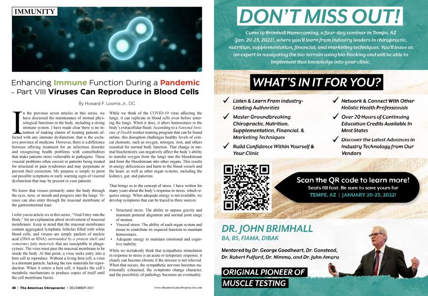
I refer you to article six in this series, “Viral Entry into the Body,” for an explanation about involvement of mucosal membranes. Keep in mind that the mucosal membranes contain aggregated lymphatic follicles filled with white blood cells, and viruses are simply packets of nucleic acid (DNA or RNA) surrounded by a protein shell and sometimes fatty materials that are susceptible to phagocytosis. The virus must pass the mucosal membrane to be inside the body. At that point, a virus seeks entry into a host cell to reproduce. Without a living host cell, a virus is a dormant particle, lacking the raw materials for reproduction. When it enters a host cell, it hijacks the cell’s metabolic mechanisms to produce copies of itself until the cell membrane bursts.
While we think of the COVID-19 virus affecting the lungs, it can replicate in blood cells even before entering the lungs. When it does, it alters homeostasis in the body’s extracellular fluids. According to a National Institute of Health worker training program that can be found online, this disruption challenges healthy levels of critical elements, such as oxygen, nitrogen, iron, and others essential for normal body function. That change in normal biochemistry can negatively affect the body’s ability to transfer oxygen from the lungs into the bloodstream and from the bloodstream into other organs. This results in energy deficiencies and harm to the blood vessels and the heart, as well as other organ systems, including the kidneys, gut, and pancreas.
That brings us to the concept of stress. I have written for many years about the body’s response to stress, which requires energy. When adequate energy is not available, we develop symptoms that can be traced to three sources:
• Structural stress: The ability to oppose gravity and maintain postural alignment and normal joint range of motion.
• Visceral stress: The ability of each organ system and tissue to contribute its required function to maintain homeostasis.
• Adequate energy to maintain emotional and cognitive stability.
While we mistakenly think that sympathetic stimulation in response to stress is an acute or temporary response, it clearly can become chronic if the stressor is not relieved. When that occurs, the sympathetic nervous becomes nutritionally exhausted, the symptoms change character, and the possibility of pathology becomes an eventuality.
The Body’s First Step in Every Pathological Process is Inflammation
The simplest and most fundamental reaction of the body to tissue irritation is inflammation. In fact, inflammation is defined as the local reaction of the body to irritation. From this definition, it is evident that any irritant may act as a cause of inflammation, and a full list of causes would include every known irritant.
Inflammation is the most common and the most carefully studied of all the fascinating changes, which the body undergoes as the result of disease. Its history is the history of pathology.
There are no new body processes in disease, only symptoms produced by normal homeostatic processes that are going too fast, too slow, or are otherwise out of time with need functions.
As you know, the word inflammation literally means “a burning.” The condition was studied clinically hundreds of years before any true insight was obtained about the inner pathological meaning of the process. In the first century A.D., Aulus Cornelius Celsus named the famous cardinal signs of inflammation as calor, rubor, tumor, and dolor in words that have subsequently become celebrated
(heat/fever, redness, swelling, and pain). Loss of function (muscle contraction and loss of range of motion) was added to the definition of inflammation by Rudolph Virchow in the nineteenth century. Virchow’s text, Cellular Pathology, laid the foundations of modern pathology. Virchow also observed that a white blood cell count would rise following the ingestion of food, termed digestive leukocytosis.
In the course of time, it became evident that these cardinal signs were the outward expression of vascular changes. In the middle of the nineteenth century, Julius Friedrich Cohnheim applied the experimental method to the study of inflammation and showed the all-important part played by blood vessels in the process. It remained for Elie Metchnikoff in 1892 to demonstrate that the central theme of inflammation was the reaction of the wandering mesodermal cells against the irritant.
So we begin to see that there is “nothing new under the sun,” except SARS, COVID-19, and other viruses, many yet to come. However, the body will respond to them as it has to every other irritant, infestation, and infection for centuries.
I would like to close this article by calling to mind some of the basics of human development that we are all taught regardless of professional specialization. What are the mesenchymal wandering cells that Metchnikoff studied before the dawn of the twentieth century, and where do they come from?
Connective tissue (CT) is the supportive framework of an organ or gland and serves to connect and support the other primary tissue types. It is derived from embryonic mesenchyme. Classification of CT is primarily based on the composition and organization of the extracellular matrix and its functions. It is tissue-specific and composed of protein fibers (collagen, reticular, and elastic) and ground substance (amorphous gel-like substance). Unlike other tissue types, which are composed primarily of cells, CT consists of only a few dispersed, inconspicuous cells within a prominent extracellular matrix.
1. Fibroblasts are the principal resident cells of connective tissue and are responsible for its synthesis and maintenance.
2. Within connective tissue, several types of cells, primarily leukocytes (white blood cells), can be found; some are long-lived in the tissue (resident cells) while others are transient and short-lived (wandering cells).
And there you have it. The body’s defense mechanisms against any irritant, infection, or infestation are found in its structural tissues. The body is capable of producing an inflammatory response, which features localized heat/ fever, redness, swelling, pain, and involuntary muscle contractions responsible for loss of joint range of motion. All it needs to remain healthy is someone to identify the source of the stress, the tissues affected, to reduce or remove it, and to supply adequate nourishment to restore normal energy production.
As always, I invite your comments and questions. You can email me at [email protected].
Dr. Howard F. Loomis Jr. has an extensive background in food enzyme nutrition. He is the president of the Food Enzyme Institute. The Food Enzyme Institute offers in-person and online seminars to healthcare practitioners around the country. Dr. Loomis published Enzymes: The Key to Health in 1999, The Enzyme Advantage in 2015, and The Enzyme Advantage for Women in 2016. His latest book, What Is Your Nutritional Deficiency?: Find It, Fix It, and Feel Better! was published in 2019. Contact info: 478 Commerce Dr. Suite 201, Madison, Wl 53719, [email protected], 800-662-2630.
Reference
1. Poor Gut Health Connected to Severe COVID, American Society of Microbiology, January 12, 2021.