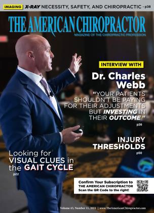
Jenkins et al. got it right when they reported, “The use of routine spinal X-rays within chiropractic has a contentious history. Elements of the profession advocate for the need for routine spinal X-rays to improve patient management, whereas other chiropractors advocate using spinal X-rays only when endorsed by current imaging guidelines.” (pg. 1) Herein lies the problem when practitioners clinically need an X-ray to conclude an accurate diagnosis, prognosis, and treatment plan and are investigated or denied reimbursement for an essential patient-centered tool. Who is creating those guidelines? The answer in the chiropractic profession is political organizations, insurance companies, and third-party administrators. Financially driven agendas could further complicate the issue. Are those in politics making these guidelines while being financially compensated by insurance carriers and third-party administrators, causing serious “conflicts of interest” at the patients’ and practitioners’ expense? Does that create a public safety issue? Those are topics for a different conversation; this article focuses on the necessity and safety of using X-ray in clinical chiropractic.
Necessity
Screening in medicine is a strategy used to look for “as-yet-unrecognized conditions” or “risk markers.” We do not take X-rays to screen patients. Instead, X-rays are used as part of the spinal examination that cannot be achieved from a clinical evaluation. Approximately 38.4% of men and women will be diagnosed with cancer at some point during their lifetimes (based on 2013-2015 data). What is the risk of treating (chiropractic spinal adjustment) a patient with undiagnosed and diagnosed (metastatic) cancer, various types of arthritides, aneurysms, osteomyelitis, ankylosing spondylitis/diffused idiopathic skeletal hypertrophy (DISH), osteophytes abutting critical neurological elements, pathological stenosis, medical subluxation, antero, and posterolestheisis, or any other condition that can increase the risk of fracture or neurological damage? The risks are numerous. Perhaps these might be acceptable losses for the carriers or political organizations, but not treating doctors. Routine use of X-rays as part of our clinical examination is a patient-centered approach, and it’s no different than segmental radiographic analysis based on patient presentation, past medical history, and physical examination findings.
Where Jenkins et al. got it wrong was when they reported, “Current evidence supports the use of spinal X-rays (in chiropractic) only in the diagnosis of trauma and spondyloarthropathy, and in the assessment of progressive spinal structural deformities, such as adolescent idiopathic scoliosis.” (pg. 1) They missed most potential pathologies and anomalies commonly diagnosed in chiropractic offices that dramatically change the diagnosis, prognosis, and treatment plan. Beyond anatomical (fracture, tumor, and infection) pathology, too many are ignoring biomechanical pathology.
B.J. and D.D. Palmer discussed spinal biomechanical pathology using the terminology “vertebral subluxation,” creating negative “vitalistic” sequela to the human body. White and Panjabi from Yale University published extensively in the 1970s and beyond on spinal kinematics, detailing normal versus pathological movement of the spine. Their research detailed biomechanical pathology and the significance of segmental pathology versus regional assessment.
A chiropractic office would have to take or order 71 lumbar X-rays to reach a level of radiation to be considered unsafe by any standard based on the scientific evidence."...
B.J. and D.D. Palmer, along with White and Panjabi’s understanding and research, has been reflected in chiropractic training, which has endured in our colleges today with teaching the use of X-ray as a tool to diagnose biomechanical pathology. Every chiropractor has been trained atlas left, 2 X PIEX (posterior, inferior, external rotation), or ASIN (anterior, superior, internal rotation) on the left. If you are a licensed chiropractor, you will understand those acronyms and, based on the evidence in the literature, cannot conclude accurate listings by palpation alone.
Seffinger et al., Troyanovich et al., and Bialosky et al. reported that palpation for position and movement faults had demonstrated poor reliability, suggesting an inability to accurately determine a specific area requiring manual therapy. Seffinger et al. went on to definitively state, “Given that the majority of palpatory tests studied, regardless of the study conditions, demonstrated low reliability, one has to question whether the palpatory tests are indeed measuring what they are intending to measure. That is to say, is there content validity of these tests? Indeed, there is a paucity of research studies addressing the content validity of these procedures. If spinal palpatory procedures do not have content validity, it is unlikely they wifi be reproducible (reliable). Obviously, those spinal palpatory procedures that are invalid or unreliable should not be used to arrive at a diagnosis, plan treatment, or assess progress.” (E419)
In contrast, the reliability of X-ray in morphology, measurements, and biomechanics, as reported by Fedorak et al., has been determined accurate and reproducible in clinical chiropractic and medicine, (p. 1858) Marques et al. reported, “In lateral cervical X-rays, reliability is excellent for all parameters, except SABB, for which it is merely good. We, therefore, suggest that this valid and reliable information on accuracy should be used when assessing and interpreting a change in cervical alignment in the context of degenerative disc disease and, with reasonable certainty, any other conditions where such parameters are used.” (pg. 175)
X-Ray Safety
The safety of X-ray in health care has been based on hypothetical models from World War II. Hendee and O’Connor report, “The hypothetical, highly speculative risks are obtained from tabulations in the Biological Effects of Ionizing Radiation VII report based primarily on data from survivors of the Hiroshima and Nagasaki atomic explosions, a population greatly different from patients experiencing medical imaging. To estimate the risks at low doses delivered by medical imaging from data greater than 100 mSv acquired from the Japanese studies, the linear no-threshold model of radiation injury is used, even though considerable evidence suggests that it is an inappropriate model for risk estimation.” (pg. 313)
Tubiana, Feinendegen, Yang, and Kaminski (2009) reported, “Among humans, there is no evidence of a carcinogenic effect for acute irradiation at doses less than 100 mSv and for protracted irradiation at doses less than 500 mSv.” (pg. 17) The American College of Radiology in their February 2020 ACR Appropriateness Criteria reported, “Adverse health outcomes for radiation doses below 100 mSv are not shown by the evidence.” They also reported (see graph below) that a lumbar X-ray is 1.4 mSvlumbar X-ray, and the cervical spine is less based on bone density comparatively.
A chiropractic office would have to take or order 71 lumbar X-rays to reach a level of radiation to be considered unsafe by any standard based on the scientific evidence. That is a threshold inconsistent with any teaching or standard in chiropractic.
Hendee and O’Connor reported that no prospective epidemiologic study with nonirradiated control subjects has quantitatively demonstrated adverse effects of radiation at doses less than 100 mSv. The American Association of Physicians in Medicine reported, “Predictions of hypothetical cancer incidence and deaths in patient populations exposed to such low doses are highly speculative and should be discouraged. These predictions are harmful because they lead to sensationalists articles in the public media that cause some patients and parents to refuse medical imaging procedures, placing them at substantial risk by not receiving the clinical benefits of the prescribed procedures.”
Conclusion
Based on a patient-centered approach to the utilization of X-ray in chiropractic, dogma, politics, and financial gain have no place when clinical outcomes are at risk. There is strong evidence that a spinal biomechanical analysis is required where palpation has shown poor reliability, and an X-ray has been found to be valid and reliable. The safety of spinal diagnostic X-ray has been deemed safe when used in the context of a chiropractic office with “no adverse effects.” When a clinician deems it necessary, X-rays as part of a clinical examination to conclude an accurate diagnosis, prognosis, and treatment plan serves two primary purposes. First, it ensures no underlying anatomical pathology, clearing the way for treatment. Second, it will render an accurate biomechanical diagnosis.
The hard rule is, “If you don’t know, don’t guess.”
References
1. Jenkins, H. J., Dow me, A. S., Moore, C. S., & French, S. D. (2018). Current evidence for spinal X-ray use in the chiropractic profession: a narrative review. Chiropractic & manual therapies, 26, 48. https://doi.org/10.1186/sl2998...
2. https://enavikipedia.org/wiki/8creemngjmedicine)
3. https://www.caiKer.gov/about-c...
4. White, A.A., Panjabi, M.A. 1975 Spinal kinematics. The research status of spinal manipulative therapy. NINCDS Monograph 15, 93.
5. White, A. A., 3rd, Johnson, R. M., Panjabi, M. M., & Southwick, W. O. (1975). Biomechanical analysis of clinical stability in the cervical spine. Clinical orthopaedics and related research, (109), 85-96. https://doi.Org/10.1097/000030...
6. Bialosky, J. E., Bishop, M. I)., Price, D. I)., Robinson, M. E., & George, S. Z. (2009). The mechanisms of manual therapy in the treatment of musculoskeletal pain: a comprehensive model. Manual therapy, 14(5), 531-538. https://doi.org/10.1016f. math. 2008.09.001
7. Seffinger, M. A., Najm, W. L, Mishra, S. I., Adams, A., Dickerson, KM., Murphy, L. S., & Reinsch, S. (2004). Reliability of spinal palpation for diagnosis of back and neck pain: a systematic review of the literature. Spine, 29(19), E413-E425. https://doi. org/10.109 7/01. brs. 0000141178.9815 7.8e
8. Troyanovich S. J. (2000). Motion palpation: it’s time to accept the evidence. Journal of manipulative and physiological therapeutics, 23(7), 514. https://doi.org/10.1067/mmt.20...
9. Fedorak, C., Ashworth, N., Marshall, J., & Pauli, H. (2003). Reliability of the visual assessment of cervical and lumbar lordosis: how good are we?. Spine, 28(16), 1857-1859. https://doi. org/10.109 7/01.BRS. 0000083281.48923.BD
10. Marques, C., Granstrom, E., MacDowall, A., Moreira, N. C., Skeppholm, M., & Olerud, C. (2020). Accuracy and reliability of X-ray measurements in the cervical spine. Asian spine journal, 14(2), 169 176. https://doi.org/10.31616/asj.2...
11. Tubiana, M., Feinendegen, L. E., Yang, C., & Kaminski, J. M. (2009). The linear no-threshold relationship is inconsistent with radiation biologic and experimental data. Radiology, 251(1), 13-22. https://doi. org/10.1148 radio!. 2511080671
12. Wang, J. ACR Appropriateness Criteria Radiation Dose Assessment Introduction
13. Wang, J. ACR Appropriateness Criteria Radiation Dose Assessment Introduction
14. Hendee, W. R., & O’Connor, M. K. (2012). Radiation risks of medical imaging: separating fact from fantasy. Radiology, 264(2), 312-321. https://doi.org/10.1148/radiol. 12112678
15. American Association of Physicists in Medicine. Position statement of the American Association of Physicists in Medicine. Radiation risks from medical imaging procedures. December 2011. http://www. aapm. org .
Dr. Mark Studin is the founder of the Academy of Chiropractic and the Doctors PI Program (www.DoctorsPIProgram.com). He also teaches in both chiropractic and medical at various levels on MRI spine interpretation, spinal biomechanical engineering, spinal trauma pathology, triaging the injured and other spinal and trauma-related topics. Dr. Studin consults and creates strategic business strategies for chiropractors, medical doctors, hospitals and lawyers nationally. He can be reached at 631 -786-4253 or [email protected]
 View Full Issue


