Advanced Care of a Professional Kenyan Runner
FEATURE
CASE STUDY
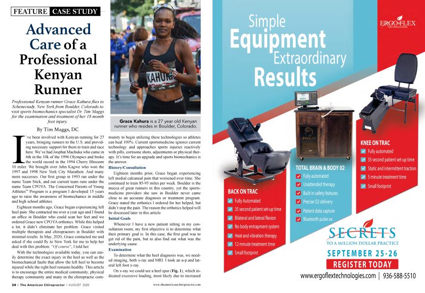
Professional Kenyan runner Grace Kahura flies to Schenectady, New York from Boulder, Colorado to visit sports biomechanics specialist Dr. Tim Maggs for the examination and treatment of her 18 month foot injury.
By
Tim Maggs
DC
I’ve been involved with Kenyan running for 27 years, bringing runners to the U.S. and providing necessary support for them to train and race here. We’ve had Josphat Machuka who came in 6th in the 10k of the 1996 Olympics and broke the world record in the 1994 Cherry Blossom 10 miler. We brought over John Kagwe who won the 1997 and 1998 New York City Marathon. And many more successes. Our first group in 1993 ran under the name Team Stick, and our current team runs under the name Team CPOYA. The Concerned Parents of Young Athletes™ Program is a program I developed 15 years ago to raise the awareness of biomechanics in middle and high school athletes.
Eighteen months ago, Grace began experiencing left heel pain. She contacted me over a year ago and I found an office in Boulder who could scan her feet and we ordered Grace new CPOYA orthotics. While this helped a lot, it didn’t eliminate her problem. Grace visited multiple therapists and chiropractors in Boulder with minimal results. In May, 2020, Grace contacted me and asked if she could fly to New York for me to help her deal with this problem. “Of course”, I told her.
With the technologies available today, you can easily determine the exact injury in the heel as well as the biomechanical faults that allow the left heel to become injured while the right heel remains healthy. This article is to encourage the entire medical community, physical therapy community and many in the chiropractic community to begin utilizing these technologies so athletes can heal 100%. Current sportsmedicine ignores current technology and approaches sports injuries reactively with pills, cortisone shots, adjustments or physical therapy. It’s time for an upgrade and sports biomechanics is the answer.
History/Consultation
Eighteen months prior, Grace began experiencing left medial calcaneal pain that worsened over time. She continued to train 85-95 miles per week. Boulder is the mecca of great runners in this country, yet the sportsmedicine providers she saw in Boulder never came close to an accurate diagnosis or treatment program. Grace stated the orthotics I ordered for her helped, but didn’t stop the pain. The reason the orthotics helped will be discussed later in this article.
Initial Goals
Whenever I have a new patient sitting in my consultation room, my first objective is to determine what their primary goal is. In this case, the first goal was to get rid of the pain, but to also find out what was the underlying cause.
Examination
To determine what the heel diagnosis was, we needed imaging, both x-ray and MRI. I took an a-p and lateral left foot x-ray.
On x-ray we could see a heel spur (Fig. 1), which indicated excessive loading, most likely due to increased miles with a collapse of the arches of the left foot. In addition to the x-ray, we would also perform a digital laser foot scan (Fig. 2) to measure the amount of space under each foot in the standing position and to see which arches of the feet have collapsed.
We also ordered a left calcaneal MRI with STIR image, which would detect fluid in the bone with greater brightness than would be found on a T2. This would tell us if bone marrow edema (bone inflammation) was present. The MRI showed bright signal at the heel spur on the STIR image (Fig. 3) and dark signal at the heel spur on the T1 view (Fig. 4). This allowed us to diagnose her injury as a stress reaction of the left calcaneus with an associated heel spur. The bigger question, however, was why the left heel became injured and the right didn’t?
Treatment
The most effective treatment for inflammation, whether it be in the soft tissue or in the bone, is cold laser therapy. My office now has 7 lasers, as most musculoskeletal conditions have an associated inflammatory phase. Cold laser also reduces pain and accelerates the healing of injuries, even fractures. We would laser Grace’s heel twice a day while she was in New York.
Biomechanical Exam
The cause of one foot becoming injured, especially in a symmetrical activity, is generally due in large part to the imbalanced biomechanics of an individual. Crooked Man (Fig. 5) shows the imbalances that originate in the feet and create a domino-like effect up the structure. With symmetrical and repetitive activity, it’s easy to understand how one specific area of the body can be overloaded, highlighting Maggs Law—When the loading of a tissue exceeds the capacity of that tissue, compensatory physiological changes occur.
All athletes fall victim to overuse injury at some point in their career because our sportsmedicine industry doesn’t identify the unique biomechanics of each athlete, or make biomechanical corrections prior to injuries occurring.
We performed our Structural Fingerprint® Exam on Grace, which involved a physical exam, 2 standing x-rays of her low back and neck and a digital laser foot scan. We then determine what the imbalances are and what we can do to reduce these imbalances, improve the healing of the current injuries and prevent future injuries.
Biomechanical Findings
When reviewing the digital foot scan (Fig. 6) it was obvious that Grace’s arches had collapsed when compared with the optimal feet. Secondly, Grace’s left foot has collapsed more than her right foot, which suggests increased loading through the left side of her body.
Over time, the foot has no choice but to collapse greater than the other foot due to overloading of the tissue.
The A-P L-S x-ray shows an unleveling of the femoral head heights (low on the left) by 8mm (Fig. 7). This may be a result of the left foot collapsing more than the right foot. Once orthotics are put in her shoes, we will re-take the A-P L-S x-ray to see what the femoral head height difference is once the arches of the feet are supported. The lateral L-S showed a normal Ferguson’s Gravity Line and Sacral Base Angle.
We continued lasering the foot (Fig. 8) twice a day, and once the orthotics arrived, we put them in Grace’s shoes and re-took the A-P L-S x-ray (Fig.9). With orthotics in her shoes, the femoral head height difference was now 1.5mm, a significant improvement as compared to not wearing orthotics. We consider 3mm and less to be normal, so no lift was needed. This explains why Grace did better with her orthotics on, but the injury still couldn’t fully heal without the laser treatments.
Conclusion
By the time Grace left New York, the foot was 60% improved as compared to when she arrived 5 days earlier. I let Grace borrow a laser so she could continue lasering her edematous heel once she returned to Colorado (Fig. 10). At the last communication with Grace, which was June 30th, she said that most days her foot is 100% but some days it’s 95%.
The key here is to be able to address both the acute injury, with an accurate diagnosis and treatment, as well as understanding the underlying biomechanical faults that have greatly contributed to the injury. Without a correction of these faults, the injury will have limited potential to fully heal and not return.
This approach to athletes as well as non-athletes is the musculoskeletal care of the future, and chiropractors are the most equipped to provide this care. We need relationships with MRI facilities, we need to put digital x-ray back in our offices, we need cold laser therapy and we need to be adjusting all of these people to keep health and happiness in all of the joints of the body. Most important, we need to be scanning every patient’s feet, as these are the foundation of the body and the governors of body alignment. When we begin doing that to every patient, we’ll realize that every human has some degree of collapse and that custom orthotics are needed if our goal is to achieve optimal alignment of the body. Grace is but one example of the success with this approach.
Dr. Tim Maggs has been in private practice for over 41 years. He has specialized in the diagnosis and treatment of sports injuries throughout his entire career. In 2004, Dr. Maggs recognized the need for improved diagnosis and treatment of sports related injuries in the middle and high school age group. It was at this time he developed the Concerned Parents of Young Athletes™ Program. Dr. Maggs launched his Structural Management® Certification Program in January 2019. He can be reached at [email protected].