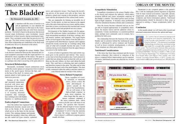
The Kidney
ORGAN OF THE MONTH
Howard F. Loomis
Jr., DC
One of the most perplexing challenges facing chiropractors today, especially those relatively new to practice, is knowing how to determine the cause of the patient’s symptoms and knowing how to convey the source of stress clearly and with confidence to the patient. I guess, in that regard, not much has changed since I started practice in the late 1960s. This month we look at the kidney, its normal functions, and how it is affected by not only the acute “fight-or-flight” response but by chronic unrelenting stress and the symptoms that arise from this organ when it cannot fulfill its responsibilities for maintaining homeostasis in the extracellular fluids.
Explaining that to patients should be quite easy because they believe all stress is mental or emotionally induced. They don’t realize that the human body is required (with no exceptions) to produce energy for three purposes:
• For structure to oppose gravity
• For visceral organs to maintain homeostasis in the extracellular fluids
• For the brain to maintain emotional and cognitive stability
There you go—only three causes for symptoms, and it gets easier because all three functions require energy.
Normal Physiological Function of the Kidney
The main function of the kidney h to filter the water-soluble substances ii the blood and discard the following:
• Waste products reabsorbed from the bowel, which must be detoxified by the liver.
Toxic substances, which are the end products of oxidative (energy-producing) metabolism.
Excess acids and bases to maintain a normal acid/ alkaline balance.
Water and salts (water/electrolyte balance) in amounts that will maintain the normal equilibrium between the extracellular and intracellular fluids.
Embryological Development of the Kidney
It is essential for practicing chiropractors and chiropractic students to understand the development of the kidney and bladder. Because these organs develop along the spine, any stress that compriseWs their normal function will cause involuntary muscle contractions along the spine and adversely influence spinal mechanics and range of motion.
All of the urinary organs develop from intermediate mesoderm, as do the urinary bladder and reproductive system. The genitourinary system develops through a series of successive phases.
The first stage begins with the appearance of the embryonic kidney (pronephros). This organ begins in the cervical region of the embryo from the fifth cervical segment to the third thoracic segment and appears on about the twenty-second day as swellings along both sides of the spine. It will continue its development from the upper thoracic area down to the lumbar spine.
A series of short evaginations from each segment grow to the posterior and inferior. They successively fuse to form the pronephric duct. Each connects to a tube that runs along the spine to drain the yolk sac, and these are the only kidneys we have until about the tenth week. They each have a collecting duct and a nephron and, therefore, connect to the aorta. The tubes end in what will become the back wall of the adult bladder. This duct is fully contained within the embryo and thus cannot excrete filtered material outside the embryo.
The second stage of development is marked by the appearance of the intermediate kidney (mesonephros), which begins as the pronephric duct elongates and proceeds downward. It induces nearby intermediate mesoderm in the thoracolumbar area to become epithelial tubules called mesonephric tubules. Each tubule ends in a capillary tuft analogous to the glomerulus and the nephron. These tubules receive a blood supply from a branch of the aorta, allowing for the filtration of blood. The filtrate flows through the tubules and is drained into a more developed continuation called the mesonephric duct or Wolffian duct. This duct extends toward the most inferior end of the embryo, ultimately attaching to the cloaca, and the embryonic kidney finally degenerates.
The cloaca is the posterior orifice in the pelvic floor that allows passage of substances from the digestive, reproductive, and urinary tracts to the outside.
During the fifth week of gestation, the mesonephric duct develops an outpouching (ureteric bud) near its attachment to the cloaca. This bud grows to the posterior and upward toward the head of the embryo. Later it forms the ureter.
The cranial end of the bud extends into the intermediate mesoderm and forms a series of branches that become the collecting duct system of the kidney. It also forms the major and minor calyces and the renal pelvis (through these organs before continuing through the renal pelvis into the ureter.)
Involuntary Muscle Contractions Associated with Kidney Stress
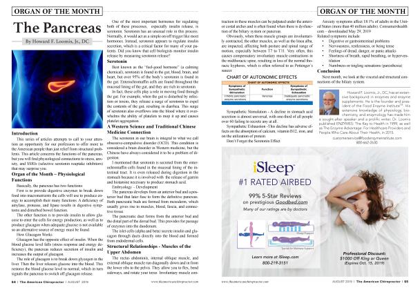
Clearly, it can be seen that any stress, be it structural, visceral, or emotional, can produce involuntary muscle contractions along the spine from the third cervical segment downward. More specifically the muscles involving dorsolumbar extension and lateral side bending are primarily affected. They are always involved to some extent in cases of lower back pain. So is the source of stress structural or visceral (kidney)? In my clinical experience, at least one-third of nontraumatic low back pain originated from visceral stress involving the kidney. The kidney receives its postganglionic sympathetic nerve supply from T10 to T12 and the renal plexus, which is located around the renal artery.
More specifically the muscles involving dorsolumbar extension and lateral side bending are primarily affected.
Involuntary muscle contractions can be felt under the lower ribs, in the back, and close to the spine. They are traceable along the bottom of the lower rib to the spine and downward to the pelvis. It should be noted that it is quite common for one kidney to be functionally dominant and for involuntary muscle contraction may be found on only one side.
Conclusion
Sympathetic visceral branches from the tenth, eleventh, and twelfth thoracic nerves exert control over the amount of blood flowing through the kidneys. Both temporary and continued stress causes vasoconstriction of the blood vessels resulting in less blood being filtered by the kidneys and reducing its efficiency at clearing unwanted water-soluble substances, such as allergens and metabolic wastes. In addition to muscular discomfort in the lower back, the patient will often complain of nausea and airborne allergies, such as hay fever. A late sign of kidney stress is seen as dark circles under the eyes.
Next month, we’ll look at visceral and structural stress and their effect on the mucosal linings.
Howard F. Loomis, Jr., DC, has an extensive background in enzymes and enzyme supplements. He is the founder and president of the Food Enzyme Institute™. His extensive knowledge of physiology, biochemistry, and enzymology has made him a sought-after speaker and a prolific writer. Dr. Loomis published ENZYMES: The Key to Health in 1999, as well as The Enzyme Advantage: For Healthcare Providers and People Who Care About Their Health, in 2015. For more informatiom, email [email protected] or call 800662-2630.