Pelvic Posture and Lower Extremity Balance
FEATURE
John K. Hyland
DC, DACBR, DABCO, CSCS
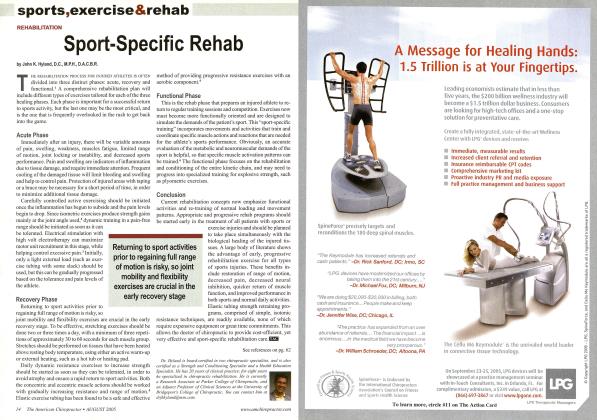
The postural alignment of the pelvis and its function during daily activities are both strongly influenced by the balance (or lack thereof) of the lower extremities. The legs affect the pelvis in three major ways. First, they provide considerable structural support—any substantial deficit will inevitably affect pelvic posture. Next, gait interactions create the vitally important interplay between the movements of the lower extremities and the corresponding repetitive motions occurring from the pelvis into the spine. Finally, proprioceptive input from nerve endings in the joints and muscles of the lower extremities provides a large amount of the information needed for the integration of pelvic and spinal postural reflexes.
We now know that a smooth, symmetrical gait has a significant effect on the pelvis and is closely tied to proper vertebral function.1 During walking, there is a fine interplay between the movements of the feet, hips, and pelvis. Orthotics and shoe lifts can either improve or interfere with the function of the pelvis.
Structural Support
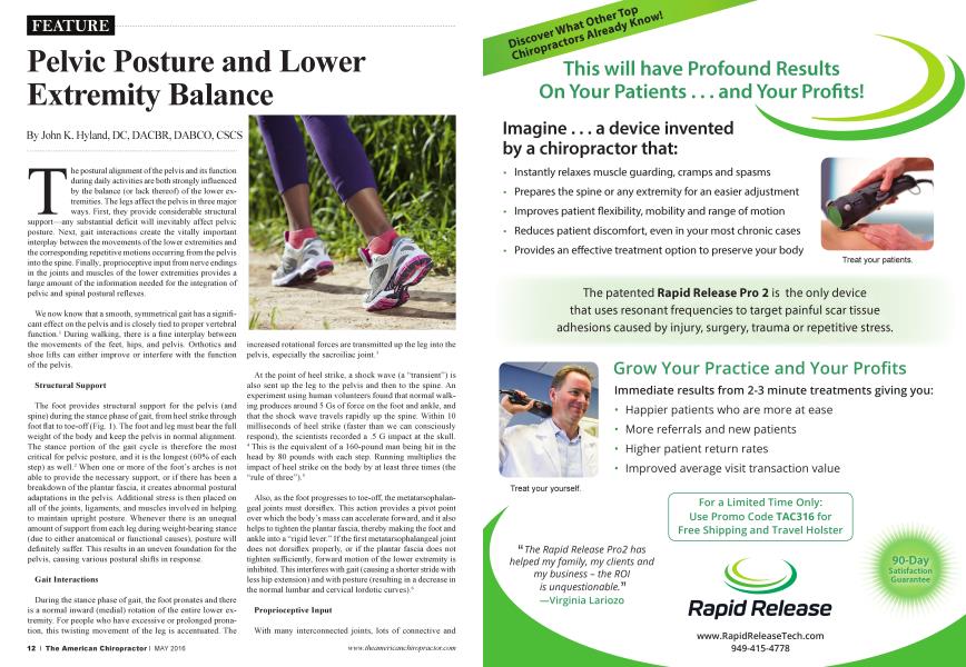
The foot provides structural support for the pelvis (and spine) during the stance phase of gait, from heel strike through foot flat to toe-off (Fig. 1). The foot and leg must bear the full weight of the body and keep the pelvis in normal alignment. The stance portion of the gait cycle is therefore the most critical for pelvic posture, and it is the longest (60% of each step) as well.2 When one or more of the foot’s arches is not able to provide the necessary support, or if there has been a breakdown of the plantar fascia, it creates abnormal postural adaptations in the pelvis. Additional stress is then placed on all of the joints, ligaments, and muscles involved in helping to maintain upright posture. Whenever there is an unequal amount of support from each leg during weight-bearing stance (due to either anatomical or functional causes), posture will definitely suffer. This results in an uneven foundation for the pelvis, causing various postural shifts in response.
Gait Interactions
During the stance phase of gait, the foot pronates and there is a normal inward (medial) rotation of the entire lower extremity. For people who have excessive or prolonged pronation, this twisting movement of the leg is accentuated. The
increased rotational forces are transmitted up the leg into the pelvis, especially the sacroiliac joint.3
At the point of heel strike, a shock wave (a “transient”) is also sent up the leg to the pelvis and then to the spine. An experiment using human volunteers found that normal walking produces around 5 Gs of force on the foot and ankle, and that the shock wave travels rapidly up the spine. Within 10 milliseconds of heel strike (faster than we can consciously respond), the scientists recorded a .5 G impact at the skull. 4 This is the equivalent of a 160-pound man being hit in the head by 80 pounds with each step. Running multiplies the impact of heel strike on the body by at least three times (the “rule of three”).5
Also, as the foot progresses to toe-off, the metatarsophalangeal joints must dorsiflex. This action provides a pivot point over which the body’s mass can accelerate forward, and it also helps to tighten the plantar fascia, thereby making the foot and ankle into a “rigid lever.” If the first metatarsophalangeal joint does not dorsiflex properly, or if the plantai' fascia does not tighten sufficiently, forward motion of the lower extremity is inhibited. This interferes with gait (causing a shorter stride with less hip extension) and with posture (resulting in a decrease in the normal lumbar and cervical lordotic curves).6
Proprioceptive Input
With many interconnected joints, lots of connective and
articular tissues, and both intrinsic and extrinsic muscles, the lower extremities are very well supplied with proprioceptive nerve endings. Mechanoreceptors in the foot, ankle, and knee joints, along with the muscle spindles of the foot and lower leg muscles, are responsible for the positive support reflexes and a variety of automatic reflexive reactions.7 The position receptors in the lower extremities, pelvis, and spine (and especially the neck/head-righting reflexes) must coordinate smoothly in order to maintain postural equilibrium. Difficulty in achieving or keeping optimal postural alignment, or problems with excessive postural sway, are frequently caused by inaccurate information sent by spindle sensors in chronically strained muscles or by joint mechanoreceptors in the lower extremities.8
In addition, much of the neurological coordination of the body is based on a balanced, rhythmic lower extremity movement and gait. The “cross crawl” pattern organizes many fundamental musculoskeletal functions at the spinal cord level, permitting smooth performance of daily physical activities without the need for conscious thinking or planning. This includes factors such as balance, stability, and center of gravity.9 When one or both feet spend too much time in pronation, many of the muscles throughout the body (and around the spine) don’t turn on and shut off in proper sequence. This raises the work effort for all activities and even increases the amount of oxygen consumed during normal walking.10
Conclusion
Lack of structural support, gait problems, and altered sensory input from the lower extremities will interfere with posture generally, but will also frequently cause specific, local biomechanical dysfunction of the pelvis. Custom-made orthotics that support the feet and provide biomechanical control during gait can reduce many of the forces previously described. Excessive stress on the pelvis from imbalances in the lower extremities can destabilize the spine and pelvis. If either the foot/ankle complex, the knee joint, or any part of the lower extremity is not functioning correctly during the stance phase of gait, abnormal strain will be transmitted to the pelvis with every step. This is just one of the ways in which providing proper orthotic support can help to alleviate many chronic pelvic conditions.
References
1. Yekutiel MR The role of vertebral movement in gait: Implications for manual therapy. J Man Manip Ther 1994; 2:22-27.
2. Magee DJ. Orthopedic Physical Assessment. Philadelphia: WB Saunders; 1987. 368.
3. Botte RR. An interpretation of the pronation syndrome and foot types of patients with low back pain. JARA 1981; 71:243-253.
4. Light LH, McLellan GE, Klenerman L. Skeletal transients on heel strike in normal walking with differentfootwear. JBiomech 1980; 13:477-480.
5. Subotnick SI. Sports Medicine of the Lower Extremity. New York: Churchill Livingstone; 1989. 67.
6. DananbergHJ. Gait style as an etiology to chronic postural pain. Part II: Postural compensatory process. J Am Podiatr Med Assoc 1993; 83:615-624.
7. Freeman MAR, Wyke BD. Articular contributions to limb muscle reflexes. J Physiol 1964; 171:20.
8. McPartland JM, Brode ur RR, Hallgren RC. Chronic neck pain, standing balance, and suboccipital muscle atrophy—a pilotstudy. J Manip Physiol Therap 1997; 20:24-29.
9. Horak FB, Nashner EM. Central programming of postural movements: Adaptation to altered support surface configuration. J Neurophysiology 1986; 55:1369-1381.
10. Otman S, et al. Energy cost of walking with flat feet. Prosthet and Orthot Inti 1988; 12:73-76.
A 1980 graduate of Logan College of Chiropractic, Dr. John Hyland practiced for more than 20 years in Colorado. In addition to his specialty board certifications in chiropractic orthopedics (DABCO) and radiology (DACBR), Dr. Hyland is nationally certified as a strength and conditioning specialist (CSCS) and a health education specialist (CHES). He has consulted chiropractors in the concepts and procedures of spinal rehabilitation and wellness exercise.