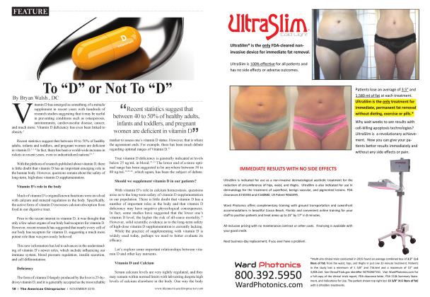
Plantar Fasciitis
ORTHOTICS
John K. Hyland
DC, DACBR, DABCO, CSCS
The classic presentation of a patient with plantar fasciitis is with “a sharp heel pain that radiates along the bottom of the inside of the foot. The pain is often worse when getting out of bed in the morning.”1 This can occur with runners or other athletes who repetitively land on the foot. Another susceptible group is middle-aged people who have spent a lot of time on their feet, either at work or during family and home care. More rarely, the fascia becomes inflamed after a single traumatic event, such as landing wrong after a jump or running a long hill. The good news is that the vast majority (95%) will respond to conservative care, and not require surgery.2 Proper treatment is necessary, though, both to ensure continued participation in sports and daily activities, and to avoid chronic damage. The plantai' fascia is the major structure that supports and maintains the arched alignment of the foot.3 This aponeurosis functions as a “bowstring” to hold up the longitudinal arch.
Pathology
Plantar fasciitis develops when repetitive weight-bearing stress irritates and inflames the tough connective tissues along the bottom of the foot. High levels of strain stimulate the aponeurosis to try to heal and strengthen. When the biomechanical strain continues, it overwhelms the body’s repair capacity, and the ligaments begin to fail. This tear/repair process causes the chronic, variable symptoms that eventually can become unbearable for some patients.
Since the plantai' fascia inserts into the base of the calcaneus, the chronic pull and inflammation can stimulate the deposition of calcium, resulting in a classic heel spur seen on a lateral radiograph. Unfortunately, there is no correlation between the presence of a heel spur and plantai' fasciitis, since many heel spurs ai e clinically silent, and most cases of plantai' fasciitis do not demonstrate a calcaneal spur.4
Examination
Biomechanical evaluation may find either excessive pronation or supination. The flatter, hyperpronating foot overstretches the bowstring function of the plantai' fascia, while the high-arched, rigid foot places excessive tension on the plantai' aponeurosis. In either case, the combination of improper foot biomechanics and excessive strain causes the connective tissue to become inflamed. A careful assessment of the weight-bearing aligmnent of the lower extremities is helpful, since many patients will have functional imbalances up the kinetic chain, into the pelvis and spine.
Direct palpation of the plantai' fascia will demonstrate discrete painful areas, most commonly at the insertion on the anteromedial calcaneus.5 Fibrotic thickenings aie frequently felt—these ai e remnants of the repetitive tear and repair process. With the
foot relaxed, grasp the toes and gently pull them up into passive dorsiflexion. Since this maneuver stretches the irritated plantai' aponeurosis, it is frequently quite painful, which is an obviously positive objective sign.
Treatment
Acute Relief
Taping - Temporary support for the strained plantar fascia can be provided with figure-eight taping or with low-dye strapping.
Restricted activity - Repetitive and straining activities should be strictly limited, initially. Immobilization is not recommended.
Cryotherapy - Ice massage and/or cold packs help reduce pain and inflammation.
Healing
Ultrasound - Initially pulsed, then constant and direct (once inflammation has subsided).
Transverse friction massage - To stimulate blood flow and collagen deposition.6
Vitamin C with bioflavonoids - A natural anti-inflammatory that can speed healing.
Adjustments
Calcaneus - Reduction of calcaneus posteriority to relieve sagittal stress. Kell’s technique uses a posterior-to-anterior thrust on a table with a pelvic drop piece.7
Other foot joints - Brantingham found various areas of joint dysfunction in the tarsal and metatarsal joints in patients with plantar fasciitis.8 The navicular and first metatarsophalangeal joints aie often involved.
Orthotic Support
Orthotics for pronation - To support the arches and reduce the stress on the plantai' fascia.
Orthotics for supination - Arch support with added viscoelastic material to cushion the foot and decrease the amount of shock at heel strike.
Heel spur correction - A “divot” in the surface of the material under the heel to spread pressure away from the fascial insertion.
Rehabilitation9
Heel and foot stretching - “Runner’s stretch” for the calf and the bottom of the foot.
Intrinsic muscle strengthening - Toe curl exercises (while sitting, gather a towel on the floor up under the arch, and repeat three times).
Extrinsic muscle strengthening - Toe raises (while standing on the edge of a stair, slowly rise up on balls of feet) and ankle stabilizing series with tubing.
Conclusion
Plantar fasciitis usually responds well to focused, conservative treatment. Steroid injections and surgical release aie seldom necessary and are best avoided. One of the most important treatment methods is to reduce any tendency to pronate excessively. In addition to custom-fitted orthotics, runners should wear well-designed shoes that provide good heel stability. The use of corrective orthotics can prevent many overuse problems from developing in the lower extremities. Investigation of foot biomechanics is a good idea for all patients, but especially for
those who aie recreationally active.
References:
1. Souza TA. Differential diagnosis for the chiropractor Gaithersburg: Aspen Publications; 1997:354.
2. Baxter DE. Hie heel in sport. Clin SportsMed 1994;13:68593.
3. Huang CK, et al. Biomechanical evaluation of longitudinal arch stability. Foot Ankle 1993; 14:353-7.
4. Lapidus PW, Guidotti FP. Painful heel: report of 323 patients with 364painful heels. Clin Orthop 1965;39:178.
5. Subotnick SI. Sports medicine of the lower extremity. New York: Churchill Livingstone; 1989:237.
6. Lear L. Transverse fiction massage. Sports Med Update 1996;10:18-25.
7. Kell PM. A comparative radiologic examination for unresponsive plantar fasciitis. J Manip Physiol Therap 1994;17:329-34.
8. Brantingham JW. Examination and treatment of plantar fasciitis Chiro Technique 1992;4:75-82.
9. Kibler WB, et al. Functional rehabilitation of sports and musculoskeletal injuries. Gaithersburg, MA: Aspen Publishers; 1998:280.
A 1980 graduate of Logan College of Chiropractic, Dr John Hylandpracticed for more than 20years in Colorado. In addition to his specialty board certifications in chiropractic orthopedics (DABCO) and radiology (DACBR), Dr Hyland is nationally certified as a strength and condition-
ing specialist (CSCS) and a health education specialist (CHES). He has consulted chiropractors in the concepts and procedures of spinal rehabilitation and wellness exercise.