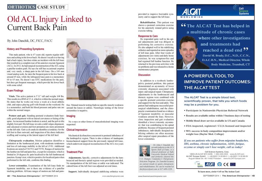
History and Presenting Symptoms This male patient, who is 47 years old. reports regular stiffness and aching in his lower back. He docsn"t recall ever having had a back injury, but docs relate an incident with his left knee that resulted in a complete tear of his anterior cniciatc ligament (ACL). In 2012. he had surgical reconstruction of the ACL using a patcllar tendon graft. He says he still has occasional crcpitus and. very rarely, a sharp pain in his left knee. On a 100 mm visual analog scale, he rates the frequent pain in his low back at around 45 mm. while the infrequent knee pain is a momentary 60 to 65 mm. He doesn't use OTC medications for the pain, but docs get frequent massages, which provide his lower back with some relief. Exam Findings Vitals. This active patient is 5" 11" tall and weighs 168 lbs. This results in a BMI of 23.4. which is within the normal range. He states that he works out twice a week at a local athletic club, and enjoys playing golf with friends on the weekend. He is a nonsmokcr. and both his blood pressure and pulse rate arc within the normal ranges. Posture and gait. Standing postural evaluation finds basically good alignment with no lateral curvatures or listing of the spine. An increased lumbar lordosis is noted, and the pelvis tilts slightly forward. His left knee reveals a mild valgus alignment, and calcancal eversion and foot pronation arc more prominent on the left side. Gait cycle analysis identifies a tendency for the left foot to flare outward, and inspection of his shoes indicates scuffing and wearing at the lateral aspect of his left heel. Chiropractic evaluation. Motion palpation identifies a limitation at the lumbosacral joint, with moderate tenderness and loss of end-range mobility to the left at L5/S1. Additional fixations arc noted at T10/T11 andT5/T6. Ranges of motion for the lumbar arc full and pain-free, except for extension, which is limited to 20 degrees by pain localized to the left lumbosacral junction. Kemps test, which is positive for localized pain when performed to the left side, confirms this finding. Lower extremities. Examination of the left knee finds no ligament instability, nor is there any evidence of a patellar tracking problem. All knee ranges of motion arc full and pain- free. Manual muscle testing finds no specific muscle weakness around the knees or ankles. Neurologic testing of the lower extremities is negative. Imaging No x-rays or other forms of musculoskclctal imaging were requested. Clinical Impression Mechanical dysfunction concurrent to postural imbalance of the lumbopclvic region. There is also evidence of inadequate biomcchanical support from the previously injured left knee, which underwent surgical reconstruction of the ACL two years ago. Treatment Plan Adjustments. Specific, corrective adjustments for the lum-bosacral and thoracic spinal regions were provided as needed. No manipulation of the left knee or ankle was indicated. The cuboid bone of the left foot was adjusted several times. Support. Individually designed stabilizing orthotics were provided to improve foot/ankle symmetry and to support the left knee. Rehabilitation. This patient was shown a postural correction exercise for his anteriorly rotated pelvis using exercise tubing. Response to Care He responded quite well to his spinal adjustments and was diligent in performing his corrective exercise. He also adapted well to his stabilizing orthotics and reported no more episodes of left knee pain. After four weeks of adjustments (six visits) and daily home exercises, he was symptom-free and had regained full lumbar function. He returned to his previous activities with no problems and was released to a home maintenance program. Discussion In addition to a textbook lumbo-pclvic postural problem, this patient demonstrated an asymmetry of lower extremity alignment associated with injury and surgical repair. Chiropractic adjustments to the lunibosacral and thoracic regions were combined with a specific postural correction exercise and support for the foot and ankle. This patient had undergone successful post-surgical rehabilitation, and the chiropractic examination found no evidence of ligament instability or muscular weakness around the knee. However, close inspection and gait evaluation identified a lower extremity asymmetry that was contributing to his spinal problems. Because of frequent postural imbalances, individually designed stabilizing orthotics are often necessary after surgical repair procedures of the lower extremities. Dr. John J. Danchik, the seventh inductee to the AC A Sports Hall of Fame, is a clinical professor at Tufts University Medical School and formerly chaired the U.S. Olympic Committee's Chiropractic Selection Program. Dr. Danchik lectures on current trends in sports chiropractic and rehabilitation. He can be reached at docforjocsid/aol.com or 617-489-1220.