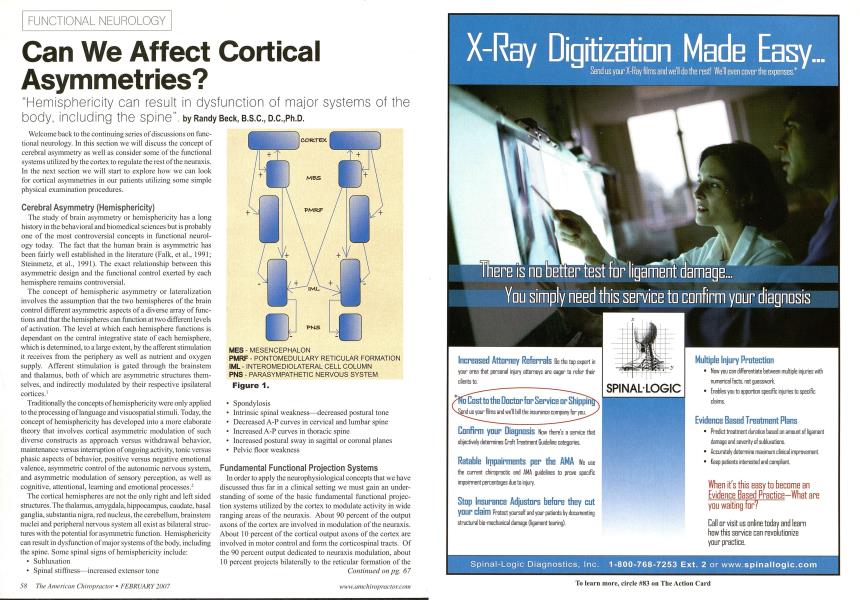
"Hemisphericity can result in dysfunction of major systems of the body, including the spine". Welcome back to the continuing series of discussions on functional neurology. In this section we will discuss the concept of cerebral asymmetry as well as consider some of the functional systems utilized by the cortex to regulate the rest of the neuraxis. In the next section we will start to explore how we can look for cortical asymmetries in our patients utilizing some simple physical examination procedures. Cerebral Asymmetry (Hemisphericity) The study of brain asymmetry or hemisphericity has a long history in the behavioral and biomedical sciences but is probably one of the most controversial concepts in functional neurology today. The fact that the human brain is asymmetric has been fairly well established in the literature (Falk, et al., 1991; Steinmetz, et al., 1991). The exact relationship between this asymmetric design and the functional control exerted by each hemisphere remains controversial. The concept of hemispheric asymmetry or lateralization involves the assumption that the two hemispheres of the brain control different asymmetric aspects of a diverse array of functions and that the hemispheres can function at two different levels of activation. The level at which each hemisphere functions is dependant on the central integrative state of each hemisphere, which is determined, to a large extent, by the afferent stimulation it receives from the periphery as well as nutrient and oxygen supply. Afferent stimulation is gated through the brainstem and thalamus, both of which are asymmetric structures themselves, and indirectly modulated by their respective ipsilateral cortices.1 Traditionally the concepts of hemisphericity were only applied to the processing of language and visuospatial stimuli. Today, the concept of hemisphericity has developed into a more elaborate theory that involves cortical asymmetric modulation of such diverse constructs as approach versus withdrawal behavior, maintenance versus interruption of ongoing activity, tonic versus phasic aspects of behavior, positive versus negative emotional valence, asymmetric control of the autonomic nervous system, and asymmetric modulation of sensory perception, as well as cognitive, attentional, learning and emotional processes.2 The cortical hemispheres are not the only right and left sided structures. The thalamus, amygdala, hippocampus, caudate, basal ganglia, substantia nigra, red nucleus, the cerebellum, brainstem nuclei and peripheral nervous system all exist as bilateral structures with the potential for asymmetric function. Hemisphericity can result in dysfunction of major systems of the body, including the spine. Some spinal signs of hemisphericity include: • Subluxation • Spinal stiffness—increased extensor tone Figure 1. • Spondylosis • Intrinsic spinal weakness—decreased postural tone • Decreased A-P curves in cervical and lumbar spine • Increased A-P curves in thoracic spine • Increased postural sway in sagittal or coronal planes • Pelvic floor weakness Fundamental Functional Projection Systems In order to apply the neurophysiological concepts that we have discussed thus far in a clinical setting we must gain an understanding of some of the basic fundamental functional projection systems utilized by the cortex to modulate activity in wide ranging areas of the neuraxis. About 90 percent of the output axons of the cortex are involved in modulation of the neuraxis. About 10 percent of the cortical output axons of the cortex are involved in motor control and form the corticospinal tracts. Of the 90 percent output dedicated to neuraxis modulation, about 10 percent projects bilaterally to the reticular formation of the Continued on pg. 67 Can We Affect Cortical Asymmetries? by Randy Beck, B.S.C., D.C,Ph.D.—Continuedfrom pg. 58 mesencephalon (MRF) and 90 percent projects ipsilaterally to the reticular formation of the pons and medulla or pontomed-ullary reticular formation (PMRF). (Fig.l, on pg. 58) The cortical projections to both the MRF and the PMRF arc excitatory in nature. The neurons in the MRF project bilaterally to excite neurons in the intermediolateral (1ML) cell columns located between Tl and L2 spinal cord levels in the grey matter of the spinal cord.' Some neurons in the PMRF project bilaterally to inhibit the neurons in the intermediolateral (IML) cell columns located between Tl and L2 spinal cord levels in the grey matter of the spinal cord, however the majority of the fibers remain ipsilateral.' These neurons in the IML form the presynaptic output neurons of the sympathetic nervous system. Neurons in the IML project to inhibit neurons in the sacral spinal cord regions that form the output neurons of the parasympathetic nervous system. Following the stimulus flow through the functional system, we can see that high cortical output results in high PMRF output which results in strong inhibition of the IML which, in turn, results in disinhibition of the sacral parasympathetic output. The bilateral excitatory output of the MRF is over-shadowed by the powerful stimulus from the cortex to the PMRF. To further illustrate the impact that an asymmetric cortical output (hemisphericity) could potentially have clinically, consider the effects of an asymmetric cortical output on the activity levels of the sympathetic and parasympathetic system on each side of the body. Autonomic asymmetries are an important indicator of cortical asymmetry, as this reflects on fuel delivery to the brain (sympathetic system) and the integrity of excitatory and inhibitory influences on sympathetic and parasympathetic function throughout the rest of the body. The PMRF has other modulatory effects in addition to modulation of the IML neurons. All of the modulatory interactions of the PMRF have clinical relevance and also include: 1) Inhibition of pain ipsilaterally, 2) Inhibition of the inhibitory interneurons which project to ventral horn cells (VHC's) ipsilaterally, which acts to facilitate muscle tone. This is another example of inhibition in the neuraxis as discussed above. 3) Inhibition of the ipsilateral anterior muscles above T6 and the posterior muscles below T6. Randy Beck, B.Sc, D.C., Ph.D., is a graduate of I Canadian Memorial Chiropractic College. He has com- f pleted postgraduate studies in Psychology, Immunology and Neurology. He is presently involved in a number of international research projects and is co-authoring a textbook on Functional Neurology. He was formerly the Dean of Chiropractic and Basic sciences and Director of Research at the New Zealand College of Chiropractic. Presently, he practices Chiropractic Functional Neumlogy at the Papahira Neurology Center and The Maungakiekie Clinic located in Auckland, New Zealand. References 1. Savic, I. Pauli, S.Thorell, J.O. Blomqvist. G. Invivo demonstration of altered benzodiazepine receptor density in patients with generalized epilepsy. J.Neuml. Neumsurg. Psychiatry: 57:797-784. 1994. 2. Davidson, R.J. and Hugdahl, K. Brain Asymmetry. Bradford, MIT press, Cambridge and London. 1995. 3. Nyberg-Hanscn, R. Sites and mode of termination of reticulospinal fibers in the cat. An experimental study with silver impregnation methods. J. Comp. Neurol. 124.74-100. 1965.1 MES - MESENCEPHALON PMRF - PONTOMEDULLARY RETICULAR FORMATION IML - INTEROMEDIOLATERAL CELL COLUMN PNS - PARASYMPATHETIC NERVOUS SYSTEM