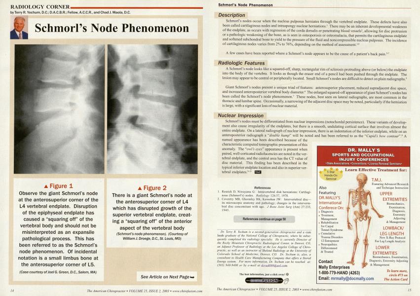
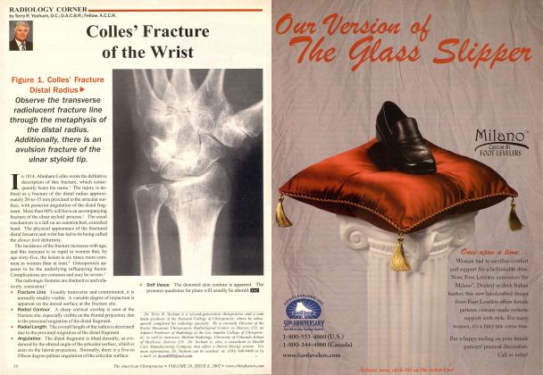
See Article on Next Page t-> Description Schmorl's nodes occur when the nucleus pulposus herniates through the vertebral endplate. These defects have also been called cartilaginous nodes and intraspongy nuclear herniations.1 There may be an inherent developmental weakness of the endplate, as occurs with regression of the corda dorsalis or penetrating blood vessels2, allowing for disc protrusion or a pathologic weakening of the bone, as is seen in osteoporosis or osteomalacia, that permits the cartilaginous endplate and softened subchondral bone to yield to the pressure of the fluid and noncompressible nucleus pulposus. The incidence of cartilaginous nodes varies from 2% to 76%, depending on the method of assessment.3-4 A few cases have been reported where a Schmorl's node appears to be the cause of a patient's back pain.5"7 Radiologic Features A Schmorl's node looks like a squared-off, sharp, rectangular rim of sclerosis protruding above (or below) the endplate into the body of the vertebra. It looks as though the eraser end of a pencil had been pushed through the endplate. The lesion may appear to be central or peripherally located. Small Schmorl's nodes are difficult to detect on plain radiographs.5 Giant Schmorl's nodes present a unique triad of features: anterosuperior placement, reduced supradjacent disc space, and increased anteroposterior vertebral body diameter.6 The enlarged squared-off appearance of giant Schmorl's nodes has : been called the Schmorl's node phenomenon.7 These nodes, best seen on lateral radiographs, are most common in the thoracic and lumbar spine. Occasionally, a narrowing of the adjacent disc space may be noted, particularly if the herniation is large, with a significant loss of nuclear material. Nuclear Impression Schmorl's nodes must be differentiated from nuclear impressions (notochordal persistence). These variants of development also cause irregularity of the endplates, but there is a smooth, undulating cortical surface that involves almost the entire endplate. On a lateral radiograph of nuclear impression, there is an indentation of the inferior endplate, while on an anteroposterior radiograph a "double hump" will be noted and has been referred to as the "Cupid's bow contour".* A named appearance has been described because of the characteristic computed tomographic presentation of this anomaly. The "owl's eyes" appearance is present when paired, well-corticated radiolucencies are noted in the vertebral endplate, and the central area has the CT value of disc material. This finding has been described in the typical inferior endplate location and also in superior vertebral endplates.9" EZS References Resnick D. Niwayama G: Intravcrtcbral disk herniations: Cartilagi nous (Schmorl's) nodes. Radiology 126:57, 1978. Coventry MB, Ghormley RK. Kernohan JW: Intervertebral disc— its microscopic anatomy and pathology; changes in the interverte bral disc concomitant with age. J Bone Joint Surg (Am) 27:233, 1945. References continue on page 50 Dr. Terry R. Yoclium is a second-generation chiropractor and a cum laude graduate of the National College of Chiropractic, where he subsequently completed his radiology specialty. He is currently Director of the Rocky Mountain Chiropractic Radiological Center, in Denver, CO, an Adjunct Professor of Radiology at the Los Angeles College of Chiropractic, as well as an instructor of Skeletal Radiology at the University of Colorado School of Medicine, Denver. CO. Dr. Yochum is, also, a consultant to Health Care Manufacturing Company that offers a Stored Energy system. For more information, Dr. Yochum can be reached at: (303) 940-9400 or by e-mail at dcradn990aol.com. The best information, just a click away! © Figure 1 Observe the giant Schmorl's node at the anterosuperior corner of the L4 vertebral endplate. Disruption of the epiphyseal endplate has caused a "squaring off" of the vertebral body and should not be misinterpreted as an expansile pathological process. This has been referred to as the Schmorl's node phenomenon. Of incidental notation is a small limbus bone at the anterosuperior corner of L5. (Case courtesy of Joel G. Green, D.C., Salem, MA) Figure 2 There is a giant Schmorl's node at the anterosuperior corner of L4 which has disrupted growth of the superior vertebral endplate, creat-ing a "squaring off" of the anterior aspect of the vertebral body (Schmorl's node phenomenon). (Courtesy of William J. Droege, D.C., St. Louis, MO)